Alternate header for print version

Datasets
Featured data set:

Description: The dataset consists of 951 tissue section scans taken from 167 H&E-stained whole slides from 18 pediatric inflammatory bowel disease (IBD) patients. Slide section scans are provided as-is without any initial image pre-processing, normalization, or cropping. Slide section scans are provided as-is without any initial image pre-processing, normalization, or cropping. Labels are provided at the patient, slide (anatomic site), and section (tissue slice) levels.


Description: Chromosomes are intracellular aggregates carrying genetic information in genes, which are major objects of study in biological cytogenetics. Chromosome screening is an important part of prenatal care. Manual identification is time consuming and costly (each image takes at least 15 minutes). We have developed an artificial intelligence (AI) model for the automatic chromosome detector based on metaphase cell images using deep learning technology.


Description: Kinome Atlas documented representative images for 456 kinases expressed in HeLa cells, and visualized by immunofluorescence staining of the epitope tag. Nuclei were stained with DAPI. Two representative views from confocal microscopy were collected for each kinase. Also, organelle markers were co-stained for some kinases to indicate specific localization.


Project ID: P20269
Description: Phenotypic profiling attempts to summarize multiparametric, feature-based analysis of cellular phenotypes of each sample so that similarities between profiles reflect similarities between samples. This image set provides a basis for testing image-based profiling methods wrt. to their ability to distinguish the effects of small molecules.


Project ID: P0000
Description: NCMIR test data for Mouse BIRN
Funding agency: NIH


Project ID: P0001
Description: Neurolucida tracing of filled Purkinje neurons
Funding agency: NIH


Project ID: P1110
Description: Investigation of morphological alterations of post-synaptic densities following an episode of transient ischemia
Funding agency: NIH
Publication: Martone M. E.; Jones Y. Z.; Young S. J. Ellisman; M. H. Zivin; J. A. and Hu B. R. Modification of postsynaptic densities following transient cerebral ischemia:a quantitative and three dimensional ultrastructural study. J. Neurosci.19:1988-1997.1999


Project ID: P1114
Description: Investigation of astrocyte morphology and the establishment of unique astrocytic domains
Funding agency: NIH
Publication: Eric A. Bushong; Maryann E. Martone; Ying Z. Jones; and Mark H. Ellisman. Protoplasmic Astrocytes in CA1 Stratum Radiatum Occupy Separate Anatomical Domains. J. Neurosci. 2002 22: 183-192.


Project ID: P1119
Description: Measurements of spine parameters using light microscopy and electron tomography
Funding agency: NIH


Project ID: P1123
Description: Confocal images
Funding agency: NIH


Project ID: P1134
Description: Actin localization in spines of CNS using eosin phalloidin photooxidation
Funding agency: NIH
Publication: Capani F; Martone ME; Deerinck TJ; Ellisman MH.Selective localization of high concentrations of F-actin in subpopulations of dendritic spines in rat central nervous system:a three-dimensional electron microscopic study.J Comp Neurol.2001 435(2):156-70.


Project ID: P1136
Description: Serial tomogram of a conventionally prepared peripheral nerve Node of Ranvier
Funding agency: NIH NINDS
Publication: Gina E.Sosinsky;Thomas J.Deerinck;Rocco Greco;Casey H. Buitenhuys;Thomas M.Bartol and Mark H. Ellisman.Development of a model for microphysiological simulations: small nodes of ranvier from peripheral nerves of mice reconstructed by electron tomography. Neuroinformatics. 2005;3(2):133-62. PMID: 15988042


Project ID: P1170
Description: Neurolucida tracing of filled Purkinje neurons
Funding agency: NIH


Project ID: P1172
Description: tests for most probable loss tomography on the 3200
Funding agency: NIH IVEM
Publication: Bouwer JC, Mackey MR, Lawrence A, Deerinck TJ, Jones YZ, Terada M, Martone ME, Peltier S, Ellisman MH. Automated most-probable loss tomography of thick selectively stained biological specimens with quantitative measurement of resolution improvement. J Struct Biol. 2004 Dec;148(3):297-306.


Project ID: P1173
Description: Confocal images
Funding agency: NIH


Project ID: P1187
Description: characterization of a mouse model of human alpha synuclein overexpressor
Funding agency: The Branfman Family Foundation
Publication: Price DL;Martone ME; Masliah MH; Ellisman MH (2003) High-resolution Large-Scale 3-D Mapping Studies of Alpha-Synuclein Immunoreactivity in Transgenic Mice Overexpressing Human Alpha-Synuclein. Society for Neuroscience Abstract.


Project ID: P1188
Description: Creation of high resolution large scale brain maps of protein expression
Funding agency: NIH


Project ID: P1194
Description: Novel specimen preparation techniques were combined with electron tomography to provide new views of synaptic structure in the cortex of the adult rat
Publication: Burette, A. C., Lesperance, T., Crum, J., Martone, M. E., Volkmann, N., Ellisman, M. and Weinberg, R. Electron tomographic analysis of synaptic ultrastructure, Journal of Comparative Neurology, in press.


Project ID: P1207
Description: Multiscale characterization of DAT KO transgenic mouse
Funding agency: NIH


Project ID: P1209
Description: In situ structures of mitochondria in rods and cones
Publication: Perkins, G.A., Ellisman, M.H. and Fox, D.A. Three-dimensional analysis of mouse rod and cone mitochondrial cristae architecture: bioenergetic and functional implications. Mol Vis. 2003 Mar 11;9:60-73.


Project ID: P1220
Description: Tomography of Caulobacter crescentus
Funding agency: Department of Energy


Project ID: P1223
Description: Reconstruction of mitochondrial associated adherens complex in the Calyx of Held synapse using electron tomography. The adherens complex is an elaborate cytoskeletal superstructure connecting a subset of mitochondria to the presynaptic membrane near active zones. Note: several additional reconstructions from this project are available from Dr. George Spirou upon request.
Funding agency: National Institutes of Health
Publication: Perkins GA, Tjong J, Brown JM, Poquiz PH, Scott RT, Kolson DR, Ellisman MH, Spirou GA. The micro-architecture of mitochondria at active zones: electron tomography reveals novel anchoring scaffolds and cristae structured for high-rate metabolism. J Neurosci. 2010 Jan 20;30(3):1015-26. PMID: 20089910


Project ID: P1226
Description: Examination of isolated mitochondria from Drosophila following infection with flock house virus (FHV-1).
Funding agency: National Institutes of Health
Publication: Kopek BG, Perkins G, Miller DJ, Ellisman MH, Ahlquist P. Three-dimensional analysis of a viral RNA replication complex reveals a virus-induced mini-organelle. PLoS Biol. 2007 Sep;5(9):e220. PMID: 17696647


Project ID: P1230
Description: Postnatal development of protoplasmic astrocytes
Funding agency: NIH
Publication: Bushong EA, Martone ME, Ellisman MH. Maturation of astrocyte morphology and the establishment of astrocyte domains during postnatal hippocampal development. Int J Dev Neurosci. 2004 Apr;22(2):73-86.


Project ID: P1231
Description: Relationship between astrocyte distribution & morpholgy and laminar boundaries in the dentate gyrus
Funding agency: National Institutes of Health
Publication: Bushong EA, Martone ME, Ellisman MH. Examination of the relationship between astrocyte morphology and laminar boundaries in the molecular layer of adult dentate gyrus. J Comp Neurol. 2003 Jul 21;462(2):241-51. PMID: 12794746


Project ID: P1233
Description: 3D investigation of T tubule structure using confocal and electron tomography from the MLP knock out mouse and human heart tissue
Publication: Hayashi, T., Martone, M. E., Yu, Z., Thor, A., Doi, M., Holst, M., Ellisman, M. H. and Hoshijima, M., Three-dimensional electron microscopy reveals new details of membrane systems for calcium signaling in the heart. J. Cell Science, 2009 Apr 1;122(Pt 7):1005-13. PMID: 19295127


Project ID: P1243
Description: This project is designed to achieve ultimate ultrastructure of animal tissues.
Funding agency: NIH
Publication: Sosinsky GE, Crum J, Jones YZ, Lanman J, Smarr B, Terada M, Martone ME, Deerinck TJ, Johnson JE, Ellisman MH. The combination of chemical fixation procedures with high pressure freezing and freeze substitution preserves highly labile tissue ultrastructure for electron tomography applications. J Struct Biol. 2007 Sep 14; PMID: 17962040


Project ID: P1576
Description: The three-dimensional morphological rearrangements for two conditions that mimic light conditions for the Antarctic summer and winter were studied in Phaeocystis antarctica Karsten
Funding agency: National Aeronautics and Space Administration
Publication: Moisan, T., Ellisman, M. H., Buitenhuys, C.W., Sosinsky, G. E., (2006) Differences in Chloroplast Ultrastructure of Phaeocystis antarctica in High and Low Light Conditions, Marine BIology, 149 (6) 1281-1290


Project ID: P1692
Description: Detailed structural investigation of the structure and distribution of synaptic densities on the spine mats from adult and embryonic ciliary ganglion neurons. Reconstructions were derived from serial electron tomography of in vivo neurons.
Funding agency: NIH
Publication: Shoop RD, Esquenazi E, Yamada N, Ellisman MH, Berg DK. Ultrastructure of a somatic spine mat for nicotinic signaling in neurons. J Neurosci. 2002 Feb 1;22(3):748-56. PMID: 11826104


Project ID: P1694
Description: Tomogram of the immunological synapse between a human cytotoxic T lymphocyte (CTL) and a target cell
Funding agency: Wellcome Trust
Publication: Jane C. Stinchcombe, Endre Majorovits, Giovanna Bossi, Stephen Fuller, Gillian M. Griffiths. Centrosome polarization delivers secretory granules to the immunological synapse. Nature 443 (2006) 462-465


Project ID: P1700
Description: Reconstruction of cortical neurons from biopsy material obtained from Alzheimers disease patients
Funding agency: National Institutes of Health
Publication: Ellisman MH, R, Ranganathan, Deerinck TJ, Young SJ, Terry R, and Mirra S: Neuronal fibrillar cytoskeleton and endomembrane system organization in Alzheimers disease, In Alteration of the Neuronal Cytoskeleton in Aging and Dementia, GP Perry (Ed,).pp, 61-73, Plenum Publ, Corp, New York, 1988.


Project ID: P1704
Description: Electron tomography was used to examine cytoskeletal-cytoskeletal, membrane-cytoskeletal, and heterologous cell connections in the paranodal region of the node of Ranvier in peripheral nerves.
Funding agency: National Institutes of Health
Publication: Perkins GA, Sosinsky GE, Ghassemzadeh S, Perez A, Jones Y, Ellisman MH. (2008) Electron tomographic analysis of cytoskeletal cross-bridges in the paranodal region of the node of Ranvier in peripheral nerves. J Struct Biol. 2008 Mar;161(3):469-80. Epub 2007 Oct 22 PMID: 18096402


Project ID: P1714
Description: Proapoptotic BH3-only proteins induce Bax/Bak-dependent mitochondrial cristae remodeling independent of cytochrome c release and Bak oligomerization
Publication: Yamaguchi, R.; Lartigue, L.; Perkins, G.; Scott, R. T.; Dixit, A.; Kushnareva, Y.; Kuwana, T.; Ellisman, M. H.; Newmeyer, D. D. Opa1-mediated cristae opening is Bax/Bak and BH3 dependent, required for apoptosis, and independent of Bak oligomerization. Molecular cell 2008, 31, 557-569. (modified)


Project ID: P1720
Description: Tomography of the virological synapse formed between an HTLV-1 infected cells and target cells.
Funding agency: Wellcome Trust
Publication: E. Majorovits, M. Nejmeddine, Y. Tanaka, G.P. Taylor, S.D. Fuller, C.R.M. Bangham: Human T-Lymphotropic Virus-1 visualized at the virological synapse by electron tomography. PLoS ONE 3: e2251, doi:10.1371/journal.pone.0002251, 2008 PMID 18509526


Project ID: P1721
Description: To characterize glial morphology and molecular markers in a mouse model of glioblastoma
Funding agency: NIH
Publication: Friedmann-Morvinski D, Bushong EA, Ke E, Soda Y, Marumoto T, Singer O, Ellisman MH, Verma IM, Dedifferentiation of neurons and astrocytes by oncogenes can induce gliomas in mice. Science. 2012 Nov 23;338(6110):1080-4. doi: 10.1126/science.1226929. [PMID:23087000]


Project ID: P1723
Description: Characterization of staining for mGluR5 glutamate receptor in animal model of Parkinsonian disorders
Funding agency: Branfman Family Foundation


Project ID: P2005
Description: Electron tomography of African Green Monkey epithelial cells infected with SARS-coronavirus
Funding agency: Leiden University Medical Center (LUMC, the Netherlands), Council for Chemical Sciences of the Netherlands Organization for Scientific Research (NOW-CW grants 700.52.306 and 700.55.002), European Commission under context of Euro-Asian SARS-DTV Network (SP22-CT-2004-511064)
Publication: Knoops K, Kikkert M, Worm SH, Zevenhoven-Dobbe JC, van der Meer Y, Koster AJ, Mommaas AM, Snijder EJ. SARS-coronavirus replication is supported by a reticulovesicular network of modified endoplasmic reticulum. PLoS Biol. 2008 6(9):e226. PMID: 18798692


Project ID: P2010
Description: Investigation of pannexins expression and distribution in the adult mouse brain.
Publication: Daniela Boassa, Phuong Nguyen, Junru Hu, Mark H. Ellisman and Gina E. Sosinsky. Pannexin2 oligomers localize in the membranes of endosomal vesicles in mammalian cells while Pannexin1 channels traffic to the plasma membrane. Frontiers in Cellular Neuroscience. fncel.2014.00468


Project ID: P2020
Description: Mutant connexins have been linked to hereditary congenital cataracts. One such mutant causes a proline-to-serine substitution at position 88 in human connexin 50 (CX50P88S). In transfected cells, CX50P88S does not form gap junctions, but localizes in cytoplasmic multilamellar structures. We studied the dynamics of formation and the stability of these structures in HeLa cells stably transfected with CX50P88S containing a tetracysteine motif appended to its C-terminus (HeLa-CX50P88S(Cys)4 cells) using a combination of light and electron microscopy.
Funding agency: National Eye Institute, National Institutes of Health
Publication: Lichtenstein A, Gaietta GM, Deerinck TJ, Crum J, Sosinsky GE, Beyer EC, Berthoud VM. The cytoplasmic accumulations of the cataract-associated mutant, Connexin50P88S, are long-lived and form in the endoplasmic reticulum Exp Eye Res. 2009 Mar;88(3):600-9. PMID: 19073179 (modified)


Project ID: P2023
Description: The Whole Brain Catalog™ is a ground-breaking, open-source, 3-D virtual environment developed by a team of researchers from UC San Diego under the Whole Brain Project™. The Catalog aims to connect members of the international neuroscience community to facilitate solutions for today's intractable challenges in brain research through cooperation and crowd sourcing. CCDB provides the backend services for very large scale image data sets on mouse brain derived from high resolution light and electron microscopy
Funding agency: Ted Waitt Family Foundation


Project ID: P2029
Description: A neuroinformatics approach to pannexin expression, localization, and interaction with other brain proteins. This research aims to examine brain tissue at high resolution and, in combination with developing neuronformatics tools, to probe for binding partners and signalling partners to investigate the pannexin "interactome". Data submissions for this project will consist of wide-scale high resolution confocal mosaic images of triple-labeled whole mouse brain sections. Pannexin 1 expression will be compared to that of pannexin 2 as well as the connexins. Other putative interaction partners of interest include the P2 purinergic receptors and components of the cytoskeleton.


Project ID: P2043
Description: To develop automated algorithms for analysis of neurite outgrowth in high content screens of cultured neurons. This data set consists of wide field epifluorescent images of cultured neurons with both cytoplasmic (phalloidin) and nuclear stains (DAPI) and a set of manual segmentations of neuronal and nuclear boundaries that can be used as benchmarking data sets for the development of segmentation algorithms.
Publication: Weimiao Yu, Hwee Kuan LEE, Srivats HARIHARAN, Wen Yu BU and Sohail AHMED. Evolving Generalized Voronoi Diagrams of Active Contours for Accurate Cellular Image Segmentation. Cytometry Part A 77A pg. 379~386, 2010.


Project ID: P2054
Description: Use of large scale imaging of the optic nerve and chiasm to characterize development of optic gliomas from neurofibromatosis-1 (Nf1) genetically-engineered mice
Funding agency: NCI and NCRR
Publication: Optic nerve dysfunction in a mouse model of neurofibromatosis-1 optic glioma. Hegedus B, Hughes FW, Garbow JR, Gianino S, Banerjee D, Kim KY, Ellisman MH, Brantley MA Jr, Gutmann DH. J Neuropathol Exp Neurol. 2009 May;68(5):542-51.


Project ID: P2061
Description: Regulation of cytoplasmic calcium is crucial both for proper neuronal function and cell survival. The concentration of Ca2+ in cytoplasm of a neuron at rest is >10,000 times lower than in the extracellular space, pointing to the importance of the transporters that extrude intracellular Ca2+. The family of plasma membrane calcium-dependent ATPases (PMCAs) represent a major component of the Ca2+ regulatory system. However, little information is available on the regional and cellular distribution of these calcium pumps. We used immunohistochemistry to investigate the distribution of each of the four PMCA isoforms in the rat brain.
Funding agency: National Institutes of Health (NS51769 EES)
Publication: Kenyon, K. A., Filoteo, A. D., Bushong, E. A., Martone, M. E., Stehler, E. E., Weinberg, R. J. and Burette, A. C. Cellular and subcellular localization of PMCA1a in the rat brain. J. Comp. Neurol. in press


Project ID: P2076
Description: During the cell cycle, gap junction communication, morphology and distribution of connexin43 (Cx43)-containing structures change dramatically. As cells round up in mitosis, the majority of Cx43 labeling is intracellular and intercellular coupling is reduced. We are interested in investigating Cx43 distributions during mitosis in exogenous expressing cells using in vivo time-lapse imaging.
Funding agency: NIH GM072881 and GM065937 awarded to Gina Sosinky, NIH GM55632 awarded to Paul Lampe
Publication: Boassa D, Solan JL, Papas A, Thornton P, Lampe PD, Sosinsky G. Trafficking and Recycling of the Connexin43 Gap Junction Protein during Mitosis. Traffic. 2010 Aug 12. in press [Epub ahead of print] PMID 20716111


Project ID: P2078
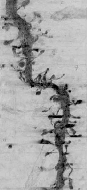
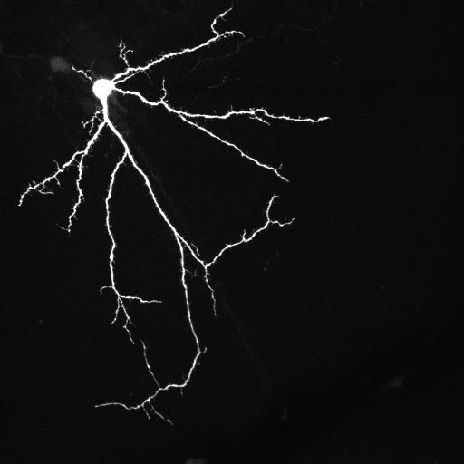
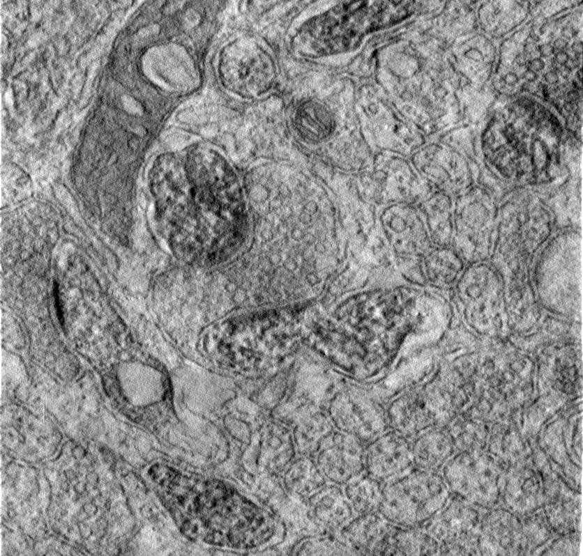
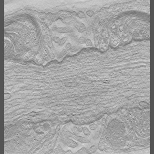
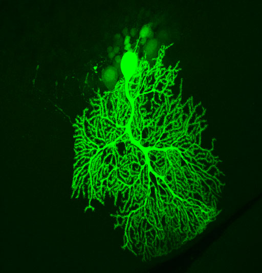
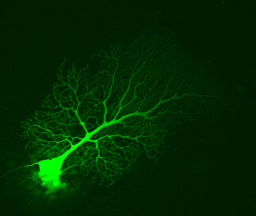
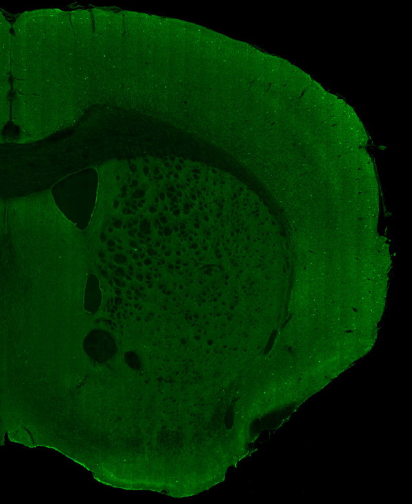
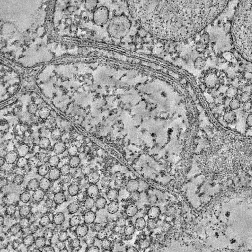
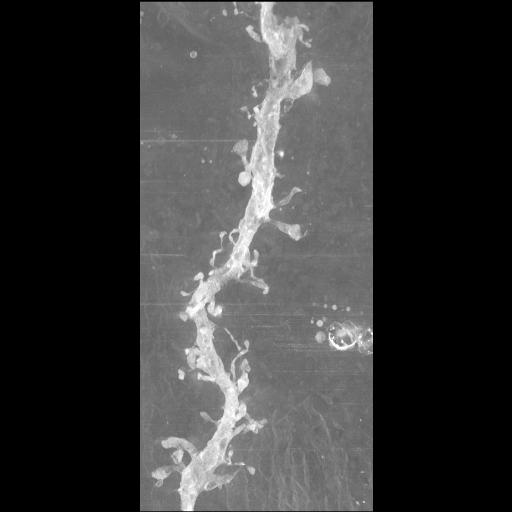
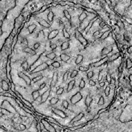
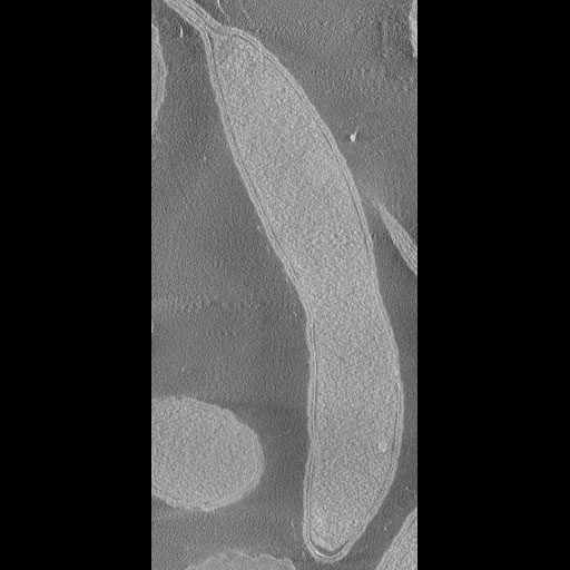
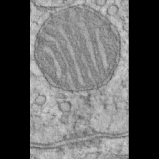
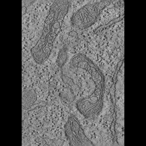
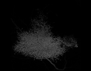
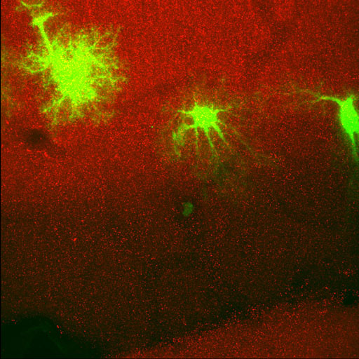
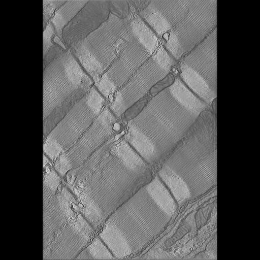
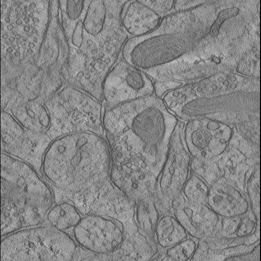
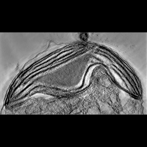
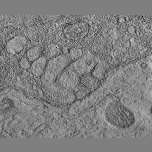
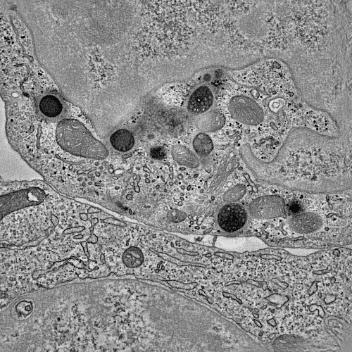
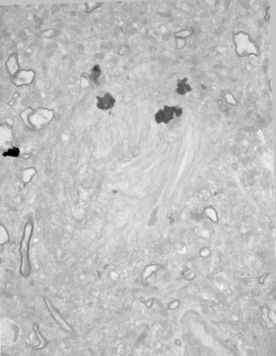
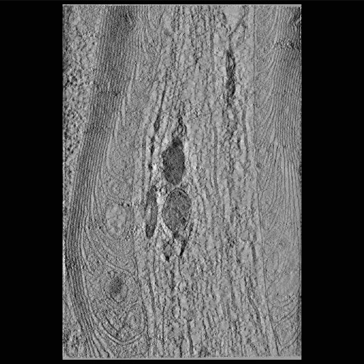
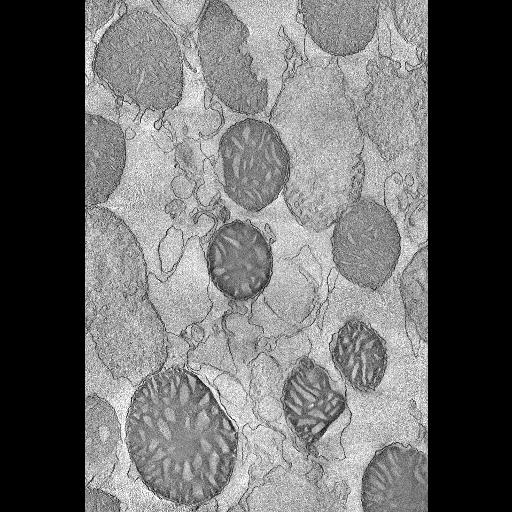
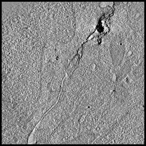
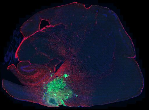
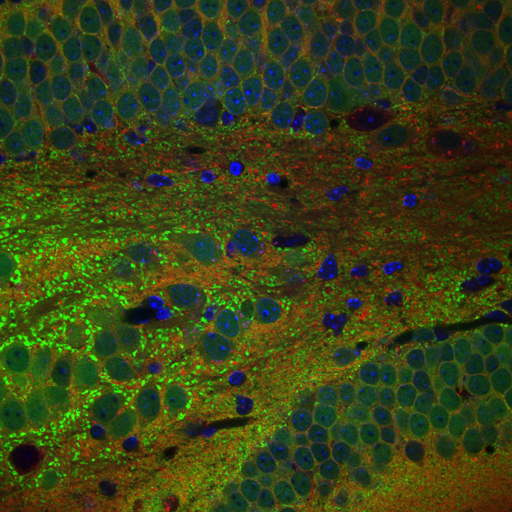
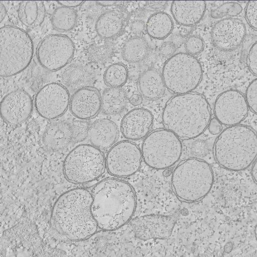
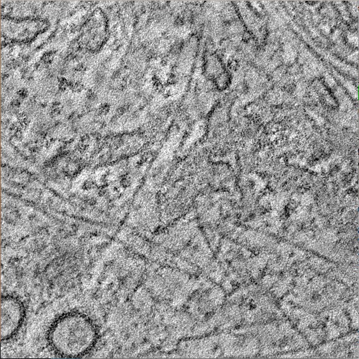
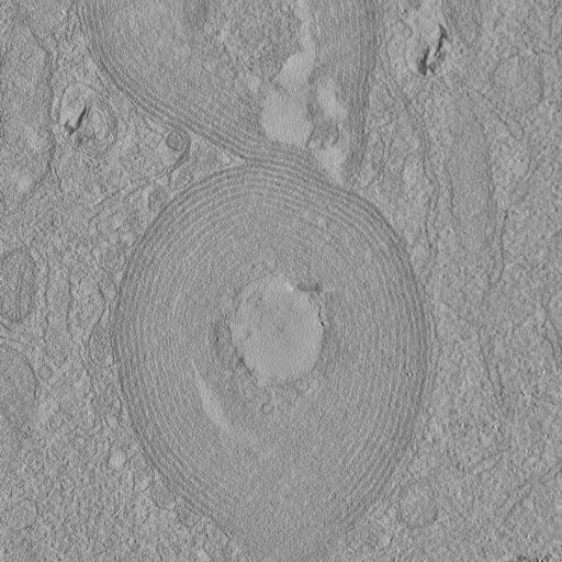
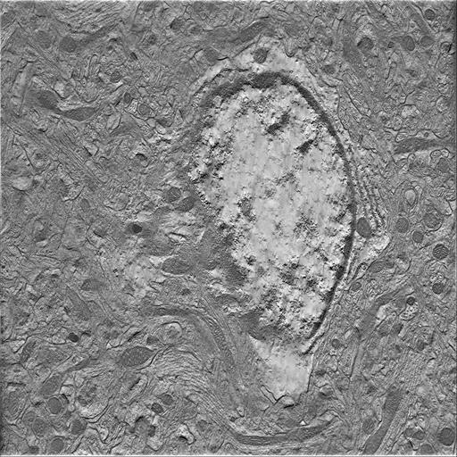
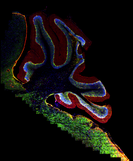
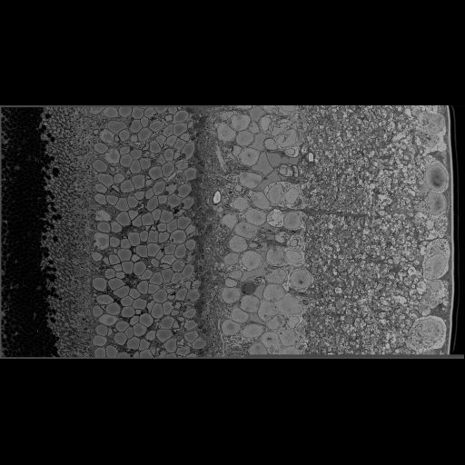
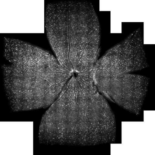
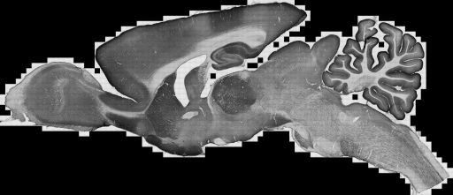
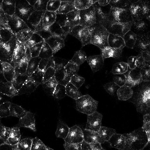
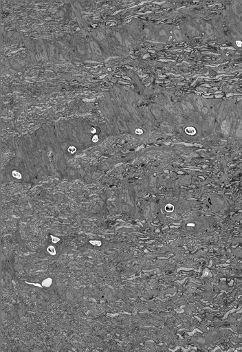
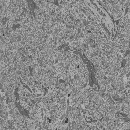
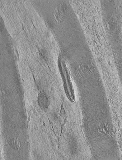
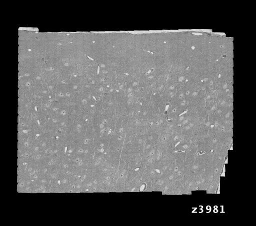
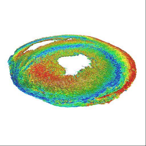
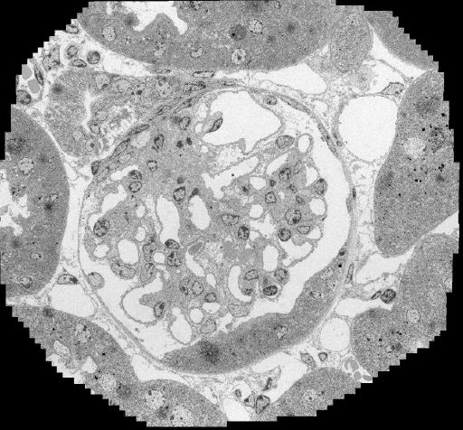
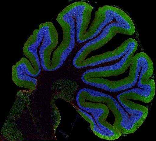
Description: 3D reconstruction using serial section scanning electron microscopy of the optic nerve head in mouse.
Funding agency: International Retina Research Foundation, and the Lasker Foundation
Publication: Nguyen JV, Soto I, Kim KY, Bushong EA, Oglesby E, Valiente-Soriano FJ, Yang Z, Davis CH, Bedont JL, Son JL, Wei JO, Buchman VL, Zack DJ, Vidal-Sanz M, Ellisman MH, Marsh-Armstrong N. Myelination transition zone astrocytes are constitutively phagocytic and have synuclein dependent reactivity in glaucoma. Proc Natl Acad Sci U S A. 2011 Jan 3. [Epub ahead of print] PubMedID:21199938


Project ID: P2080
Description: Various volumes generated on the 3View during testing or as preliminary data for other projects


Project ID: P2083
Description: Study of morphology of quantitative morphology of mitochondria in the peripheral nerve of the adult rat using 3D electron microscopy
Funding agency: National Institute of Neurological Disorders and Stroke, National Center for Research Resources, National Institute of Diabetes and Kidney Diseases, National Library of Medicine
Publication: Perkins, G.A., Ellisman, M.H. Mitochondrial configurations in peripheral nerve suggest differential ATP production. J. Struct. Biol. (2010), doi:10.1016/j.jsb.2010.06.017 http://www.ncbi.nlm.nih.gov/pubmed/20600951


Project ID: P2097
Description: Large scale serial section electron microscopic reconstruction was used to reconstruct a portion of visual cortex in mouse that had been functionally characterized for preferred stimulus orientation using two photon calcium imaging. Reconstructions showed that that inhibitory interneurons receive convergent anatomical input from nearby excitatory neurons with a broad range of preferred orientations
Funding agency: National Eye Institute
Publication: Bock DD, Lee WC, Kerlin AM, Andermann ML, Hood G, Wetzel AW, Yurgenson S, Soucy ER, Kim HS, Reid RC. Network anatomy and in vivo physiology of visual cortical neurons. Nature. 2011 Mar 10;471(7337):177-82. PMID: 21390124


Project ID: P2128
Description: High-resolution diffusion tensor MRI experiments of control and hypertenstive rats to determine fiber and sheet orientations on 17T scanner at Gainesville, Florida.


Project ID: P2133
Description: Determine the role of the endothelial glycocalyx in the glomerular filtration barrier.
Funding agency: Dutch Kidney Foundation
Publication: Martijn J.C. Dane, Bernard M. van den Berg, M. Cristina Avramut, Frank G.A. Faas, Johan van der Vlag, Angelique L.W.M.M. Rops, Raimond B.G. Ravelli, A.J. Koster, Anton Jan van Zonneveld, Hans Vink, Ton J. Rabelink. Glomerular endothelial surface layer acts as a barrier against albumin filtration. Am J Pathol, in press


Project ID: P20002
Description: A neuroinformatics approach to pannexin expression, localization, and interaction with other brain proteins. This research aims to examine brain tissue at high resolution and, in combination with developing neuronformatics tools, to probe for binding partners and signalling partners to investigate the pannexin interactome . Data submissions for this project will consist of wide-scale high resolution confocal mosaic images of triple-labeled whole mouse brain sections. Pannexin 1 expression will be compared to that of pannexin 2 as well as the connexins. Other putative interaction partners of interest include the P2 purinergic receptors and components of the cytoskeleton.
Funding agency: NIH F32GM092457
Publication: Multi-Channel Transfer Function with Dimensionality Reduction. Proc SPIE. 2010 Jan 18;7530:75300A. Kim HS, Schulze JP, Cone AC, Sosinsky GE, Martone ME. Department of Computer Science and Engineering, University of California San Diego, 9500 Gilman Drive, La Jolla, CA, USA. PMID: 20582228


Project ID: P20090
Description: Dominant missense mutations in leucine-rich repeat kinase 2 (LRRK2) are the most common genetic cause of Parkinson disease (PD) and genome wide association studies identify LRRK2 sequence variants as risk factors for sporadic PD. Intact kinase function appears critical for the toxicity of LRRK2 PD mutants, yet our understanding of how LRRK2 causes neurodegeneration is limited. We will examine how LRRK2 mutations affect the ultrastructure of cells using electron tomography.
Funding agency: NIH, Branfman Family Foundation, and Parkinson Disease Foundation.
Publication: Kett LR, Boassa D, Ho CC, Rideout HJ, Hu J, Terada M, Ellisman M, Dauer WT. LRRK2 Parkinson disease mutations enhance its microtubule association. Hum Mol Genet. 2012 Feb 15;21(4):890-9. Epub 2011 Nov 11. PMID:22080837


Project ID: P20094
Description: The goal of this project is to better understand the role of alpha-synuclein in the progression of PD by applying labeling systems developed at NCMIR to track, at multiple resolution scales, the distribution and trafficking of these proteins associated with Parkinson's disease.
Publication: Boassa D, Berlanga ML, Yang MA, Terada M, Hu J, Bushong EA, Hwang M, Masliah E, George JM, Ellisman MH. Mapping the subcellular distribution of alpha-synuclein in neurons using genetically encoded probes for correlated light and electron microscopy: Implications for Parkinson's disease pathogenesis. J Neurosci. 2013 Feb 6;33(6):2605-15. doi:10.1523/JNEUROSCI.2898-12.2013. PMID:23392688


Project ID: P20196
Description: Tomograms of specifically labeled connexin 43 (Cx43) gap junctions expressed in HEK293T. Cx43 was genetically tagged with GPF and APEX, an engineered plant peroxidase that reacts with hydrogen peroxide and diaminobenzidine to create a protein specific stain. Sections are cut perpendicular to the membrane plane (cross-section view).
Funding agency: NIH