Alternate header for print version
Advanced search
Contributors
Help
Submit
Search
menu
Cell Process
Cell Component
Cell Type
Organism
Microbial
Alzheimer's
Data Sets
Center for Research in Biological Systems
University of California, San Diego
9500 Gilman Drive
La Jolla, CA 92093-0608, USA
Voice
: (858) 534-0276
Fax
: (858) 534-7497
Email
: dorloff@ncmir.ucsd.edu
Search Results for
scanning electron microscopy
(454 results)
(Not the results you were expecting?)
(Comments)
Still Images
Video/Animation
Z-Stack
Time Series
CIL:25356
NCBI Organism Classification
Hydra viridis
Biological Process
none specified
Cellular Component
none specified
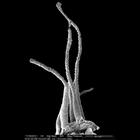
Scanning electron micrograph showing tentacles on the head of a Hydra viridis. Epithelial cells located at the base of the tentacle are in a flat, contracted state. Cell distal to the head of the H. v...
CIL:24842
NCBI Organism Classification
Hydra viridis
Biological Process
none specified
Cellular Component
none specified
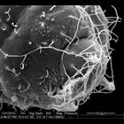
Scanning electron micrograph showing the tip of a Hydra viridis tentacle. A meshwork of triggered nematocyst cells is visible. When hooked cnidocytes on the surface of the tentacle come in contact wit...
CIL:25357
NCBI Organism Classification
Hydra viridis
Biological Process
none specified
Cellular Component
none specified
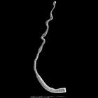
Scanning electron micrograph showing tentacles on the head of a Hydra viridis. Epithelial cells located at the base of the tentacle are in a flat, contracted state. Cell distal to the head of the H. v...
CIL:221
NCBI Organism Classification
Homo sapiens
Biological Process
gas transport
Cellular Component
none specified
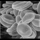
Red blood cells (erythrocytes). This image of human red blood cells obtained by scanning electron microscopy, revealing their characteristic biconcave shape.
CIL:35464
NCBI Organism Classification
Rattus norvegicus
Biological Process
none specified
Cellular Component
none specified
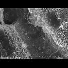
Liver sinusoid of a Brown Rat with fenestrated endothelial cells. Sinusoidal width is about 5 microns. Original magnification is 10,000x . Note the microvilli of hepatocytes in the space of Disse exte...
CIL:35465
NCBI Organism Classification
Rattus norvegicus
Biological Process
none specified
Cellular Component
none specified
Fenestrae of a Brown Rat arranged in sieve plates. Original magnification is 20,000x, a good indication of liver sinudosial endothelial cells (LESCs).
CIL:10603
NCBI Organism Classification
Leishmania mexicana
Biological Process
promastigote form
Cellular Component
none specified
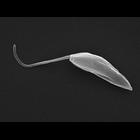
Single promastigote form Leishmania mexicana cell as seen by scanning electron microscopy. Promastigote form Leishmania mexicana (WHO strain MNYC/BZ/62/M379) were grown in M199 (10% FCS, pH 7.4, 28°C...
CIL:11091
NCBI Organism Classification
Homo sapiens
Biological Process
cell projection organization
Cellular Component
microvillus
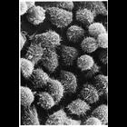
Scanning electron micrograph reveals microvilli on the surface of HeLa cells in culture. Image by Keith Porter. Figure 31 from Chapter 2 (Specializations of the Free Surface) of 'The Cell, 2nd Ed.' b...
CIL:222
NCBI Organism Classification
none specified
Biological Process
none specified
Cellular Component
plasma membrane
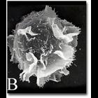
A scanning electron microscope image of an activated mast cell illustrating the convoluted topography of the cell membrane, which is populated with receptors.
CIL:41473
NCBI Organism Classification
coral
Biological Process
skeleton organization
Cellular Component
calcium skeleton
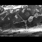
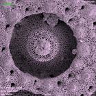
Scanning electron micrograph of the calcium skeleton of coral polyp. The sample was cleaned with bleach solution, dried and carbon coated prior to imaging. The image was collected using a secondary...
1
2
3
4
5
6
7
8
9
...
46
Next »
Results per page:
10
20
50
100