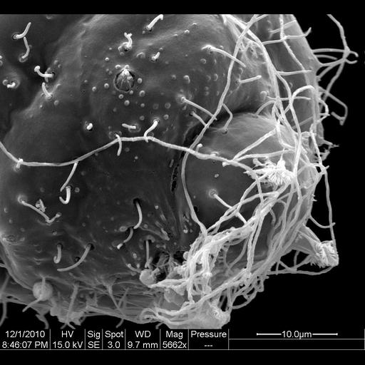
Scanning electron micrograph showing the tip of a Hydra viridis tentacle. A meshwork of triggered nematocyst cells is visible. When hooked cnidocytes on the surface of the tentacle come in contact with prey, they release a coiled nematocyst. Extension of the nematocyst thread allows the tentacle to latch onto the prey, and this signals the hypostome, or mouth, to open. Specimens were fixed in Aldehyde-osmium fixative 0.75% glutaraldehyde, 0.75% formaldehyde and 1.0% OsO4 prepared in 0.05M sodium cacodylate buffer. Fixative was buffered to pH 7.4-7.5. H. viridis were critical point dried and sputter coated with gold. Images were taken on a FEI Quanta 200 ESEM scanning electron micrograph. This sample was photographed as a montage at a magnification of 5662x and an accelerating voltage of 15kV with a 9.7mm working distance.
| Spatial Axis | Image Size | Pixel Size |
|---|---|---|
| X | 1024px | 2.12nm |
| Y | 943px | 2.12nm |