Alternate header for print version


Licensing
Public Domain: This image is in the public domain and thus free of any copyright restrictions. However, as is the norm in scientific publishing and as a matter of courtesy, any user should credit the content provider for any public or private use of this image whenever possible. Learn more
tweet  
*CIL – Cell Image Library accession number. Please use this to reference an image.

CIL:8952*  Cite 
Description

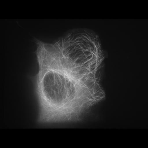
Microtubule dynamics in a U2OS (human osteosarcoma) cell. Cells were injected with Xrhodamine-tubulin and this time-lapse image series was collected on a spinning disk confocal microscope on a Nikon Ti200 using a 100X 1.4 NA Plan Apo objective with a 605x52 EM filter for 568 nm laser excitation. Images were collected with a Coolsnap HQ2 (Photometrics) with 700 ms exposure.

Biological Sources
NCBI Organism Classification
Homo sapiens
Cell Type
epithelial cell
Cell Line
U-2 OS
Cellular Component
microtubule
Biological Context
Biological Process
microtubule cytoskeleton organization
Molecular Function
microtubule plus-end binding
Attribution
Names
Yukako Nishimura
Clare M. Waterman
OTHER
Cell and Tissue Morphodynamics
Citation
Digital Object Identifier (DOI)
doi:10.7295/W9CIL8952
Archival Resource Key (ARK)
ark:/b7295/w9cil8952
Imaging
Image Type
recorded image
Image Mode
spinning disk confocal microscopy
Parameters Imaged
fluorescence emission
Source of Contrast
distribution of a specific protein
Visualization Methods
X-Rhodamine
Processing History
unprocessed raw data
Sample Preparation
Methods
living tissue
Relation To Intact Cell
dispersed cells in vitro
Dimensions
Spatial Axis Image Size Pixel Size
X 1392px 0.105µm
X 1040px 0.105µm
Time 7 seconds