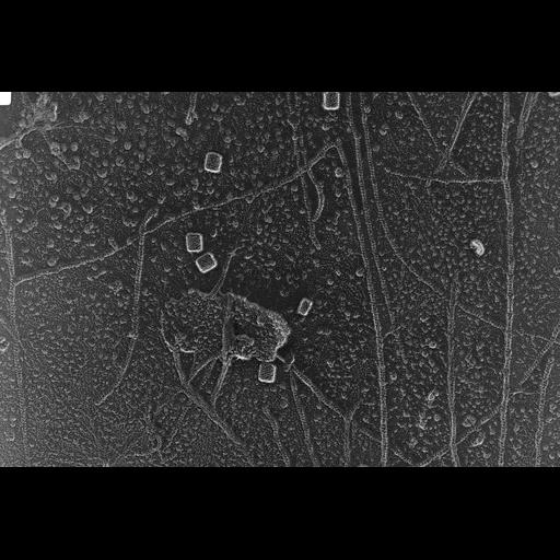
Rabbit skeletal muscle fibers were minced in a ATP-containing relaxing buffer and dissociated into thick and thin filaments by mild blending. The isolated filaments were then deposited onto mica flakes, quick-frozen by contact with a liquid helium- cooled copper block in a Heuser-type cryopress, and freeze-etched in a Balzers 400 freeze fracture machine. The sample was then rotary-replicated with Pt-C at 11 degrees and visualized in a JEOL 100CX2 transmission electron microscope operating at 100 kev. Images were recorded on Kodak 4489 film at 75,000x magnification. Stereo pairs were taken at +5 and -5 degrees. Films were scanned with a 20 micrometer pixel spacing on a Nikon Coolscan 9000ED. All the filaments seen in this micrograph are thin, actin-containing filaments. Note the striped appearance representing a helical rise due to subunit packing of g-actin monomers. A few hemocyanin molecules were included in this preparation for size calibration. RIGHT image of a stereo pair.
| Spatial Axis | Image Size | Pixel Size |
|---|---|---|
| X | 4163px | 0.00026µm |
| Y | 2790px | 0.00026µm |