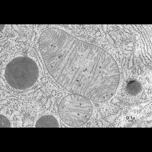
Here a mitochondrion is sliced approximately through its midplane, so both its outer membrane and the infoldings of its inner membrane are clearly seen. The organization of the inner membrane is particularly clear due to the good preservation of the cell, the thinness of the section, and the quality of this micrograph (note places where the continuity of the inner membrane with the 'cristae' that occupy the inner part of the organelle are clear). The interior of the mitochondrion is filled with a comparatively dense matrix, which includes darkly staining spherical objects that are probably crystals of calcium phosphate. Also clear is the wealth of ribosomes in the cytoplasm outside the mitochondrion. Electron micrographs like this, obtained in the early 1960s by expert microscopists, like Keith Porter and his students, opened the eyes of the scientific community to the complexity and beauty of the organization found in cells. These images are the cornerstones upon which the foundations of cell biology as a field of study were built.
Electron micrograph of a thin section of the pancreas from a bat, fixed so as to preserve the details of structure in the cytoplasm and its organelles.The pancreas of bat was excised, cut into small pieces with a sharp scalpel, and these cubes of tissue, less than 1mm on a side, were placed in buffered glutaraldehyde for several hours fixation. These samples were rinsed in buffer, post-fixed in phosphate-buffered 1% OsO4, dehydrated in a series of mixtures of water and ethanol, with increasing concentrations of the organic solvent, until the sample couple be transferred to propylene oxide, then mixtures of liquid epoxy resin with this solvent, and finally pure resin, which could be hardened by polymerization at 60 degrees Celsius for several hours. Thin sections were cut, stained with lead and uranyl acetate, the imaged in a Philips electron microscope. Micrograph was to be figure 2-35 of a revised 5th edition (unpublished) atlas by Keith Porter and Mary Bonneville (Porter KR, Bonneville MA. Fine Structure of Cells and Tissues, 4th edition. Lea & Febiger. Philadelphia, PA. 1973.). Original resource provided by Keith R Porter Archives (University of Maryland Baltimore County, Baltimore, MD). Still image; jp2; 8-Jul-65;negative.
| Spatial Axis | Image Size | Pixel Size |
|---|---|---|
| X | 4500px | 21.667µm |
| Y | 3060px | 21.667µm |