Alternate header for print version


Licensing
Public Domain: This image is in the public domain and thus free of any copyright restrictions. However, as is the norm in scientific publishing and as a matter of courtesy, any user should credit the content provider for any public or private use of this image whenever possible. Learn more
tweet  
*CIL – Cell Image Library accession number. Please use this to reference an image.

CIL:7342*  Cite 
Description

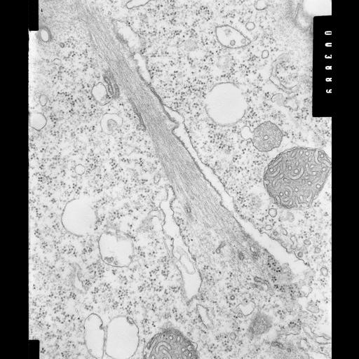
High resolution image of a myoneme of Opercularia coarctata. Such fibrous bundles are known to be composed of centrin, a filament that contracts when exposed to calcium. The ER is closely associated with the myonemes and sends specialized decorated tubules onto the surface or into the fibrous bundle. The peroxisome typically has one luminal tubule and mitochondria have tubular cristae. TEM taken on 6/13/69 by R. Allen with Philips 300 operating at 60kV. Neg. 30,200X. The raw film was scanned with an Epson Perfection V750 Pro. This image is best used for quantitative analysis. Standard glutaraldehyde fixation followed by osmium tetroxide, dehydrated in alcohol and embedded in an epoxy resin. Microtome sections prepared at approximately 75nm thickness. Additional information available at (http://www5.pbrc.hawaii.edu/allen/).

Biological Sources
NCBI Organism Classification
Opercularia [NCBITaxon:168247]
Opercularia coarctata
Cell Type
cell by organism
eukaryotic cell
Eukaryotic Protist
Ciliated Protist
Cell Line
Cambridge Culture Collection, Cambridge, England, # LB1657/1
Cellular Component
cortical actin cytoskeleton
centrin fibers
rough endoplasmic reticulum
peroxisomal membrane
peroxisomal matrix
mitochondrial inner membrane
Biological Context
Biological Process
centrin fibers
fiber targeting of rough endoplasmic reticulum
Molecular Function
intracellular ligand-gated calcium channel activity
centrin
Attribution
Names
Richard Allen
Citation
Digital Object Identifier (DOI)
doi:10.7295/W9CIL7342
Archival Resource Key (ARK)
ark:/b7295/w9cil7342
Grouping This image is part of a group.
Imaging
Image Type
transmission electron microscopy (TEM)
illumination by electrons
Image Mode
detection of electrons
film
Parameters Imaged
electron density
Source of Contrast
stain with broad specificity
Visualization Methods
stain with broad specificity
osmium tetroxide
uranyl salt
lead salt
Processing History
recorded image
film
unprocessed raw data
Data Qualifiers
raw, unprocessed data
suitable for spatial measurements
Dimensions
Spatial Axis Image Size Pixel Size
X 3886px 0.66nm
Y 4584px 0.66nm