Alternate header for print version


Licensing
Public Domain: This image is in the public domain and thus free of any copyright restrictions. However, as is the norm in scientific publishing and as a matter of courtesy, any user should credit the content provider for any public or private use of this image whenever possible. Learn more
tweet  
*CIL – Cell Image Library accession number. Please use this to reference an image.

CIL:7319*  Cite 
Description

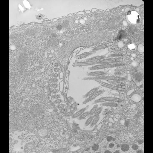
High resolution image of the buccal cavity of Halteria has oral ciliature on its left side and a paroral kinety consisting of only one row of basal bodies and cilia on its right side. A cytopharynx with microtubular lamellae arises to the right of the paroral kinety where it will continue deep into the cell as a cytopharyngeal tube accompanied by the microtubular lamellae. A thick layer of microtubules covers one side of the buccal cavity. Mitochondria are both expanded and condensed). TEM taken on 3/12/71 by R. Allen with Hitachi HU11A operating at 75kV. Neg. 9,250X. The raw film was scanned with a Nikon Coolscan 9000ED. This image is suitable for quantitative analysis. Standard glutaraldehyde fixation followed by osmium tetroxide, dehydrated in alcohol and embedded in an epoxy resin. Microtome sections prepared at approximately 75nm thickness. Additional information available at (http://www5.pbrc.hawaii.edu/allen/).

Biological Sources
NCBI Organism Classification
Halteria grandinella
Cell Type
cell by organism
eukaryotic cell
Eukaryotic Protist
Ciliated Protist
Cell Line
Carolina Biological Supply Co., NC, U.S.A.
Cellular Component
oral apparatus
cytoskeleton
ciliary rootlet
Biological Context
Biological Process
cytoskeleton organization
response to food
Attribution
Names
Richard Allen
Citation
Digital Object Identifier (DOI)
doi:10.7295/W9CIL7319
Archival Resource Key (ARK)
ark:/b7295/w9cil7319
Grouping This image is part of a group.
Imaging
Image Type
transmission electron microscopy (TEM)
illumination by electrons
Image Mode
detection of electrons
film
Parameters Imaged
electron density
Source of Contrast
stain with broad specificity
Visualization Methods
stain with broad specificity
osmium tetroxide
uranyl salt
lead salt
Processing History
recorded image
film
unprocessed raw data
Data Qualifiers
raw, unprocessed data
suitable for spatial measurements
Dimensions
Spatial Axis Image Size Pixel Size
X 4451px 1.6nm
Y 5025px 1.6nm