Alternate header for print version


Licensing
Attribution Non-Commercial Share Alike:This image is licensed under a Creative Commons Attribution, Non-Commercial Share Alike License. View License Deed | View Legal Code
tweet  
*CIL – Cell Image Library accession number. Please use this to reference an image.

CIL:54641*  Cite 
Description

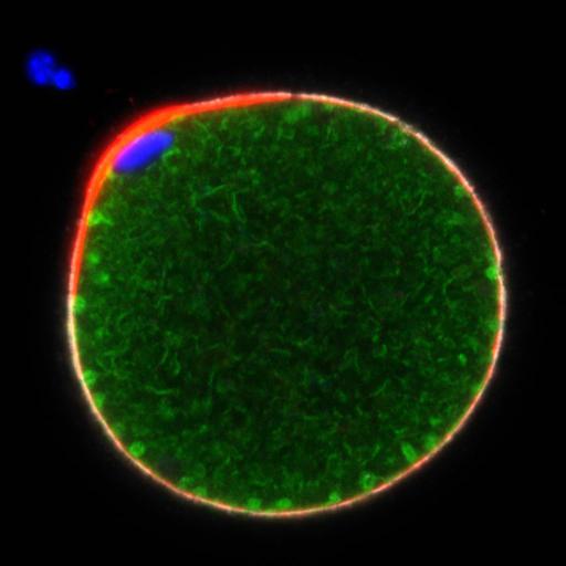
SI FIG. 1 Structure of actin layer fenestrae and relationship with plasma membrane

Technical Details

Oocytes fixed with 2% paraformaldehyde and 1% picric acid. Immunolabelled with anti-Calreticulin (green), anti-CD9(white), Alexafluor 568-phalloidin (red), and Hoechst 33342 (blue)

Biological Sources
NCBI Organism Classification
Mus musculus
Cell Type
oocyte
Cellular Component
Oocyte Cortex
Biological Context
Biological Process
Anaphase with incorporated sperm
Attribution
Names
William Kinsey
Lane Christenson
Huizhen Wang
OTHER
SI FIG. 1 from 8-18-21 egg5
Citation
Digital Object Identifier (DOI)
doi:10.7295/W9CIL54641
Archival Resource Key (ARK)
ark:/b7295/w9cil54641
Grouping This image is part of a group.
Imaging
Image Mode
whole mount confocal image
Visualization Methods
Laser Scan Confocal
PMT