Alternate header for print version


Licensing
Public Domain: This image is in the public domain and thus free of any copyright restrictions. However, as is the norm in scientific publishing and as a matter of courtesy, any user should credit the content provider for any public or private use of this image whenever possible. Learn more
tweet  
*CIL – Cell Image Library accession number. Please use this to reference an image.

CIL:50660*  Cite 
Description

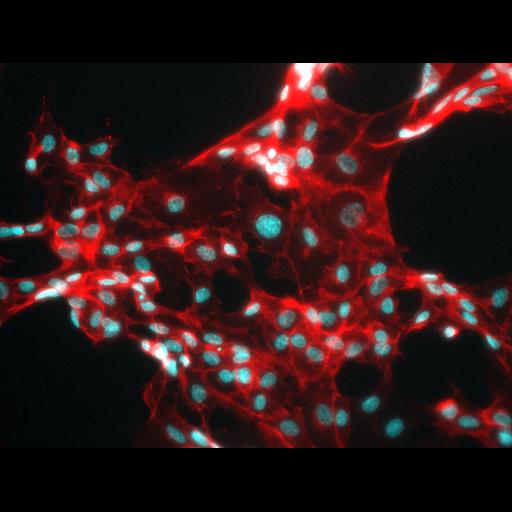
Human umbilical vein endothelial cells stained initially for nuclei with DAPI (blue) and for vascular endothelial cadherin (red).

Technical Details

Image was acquired with 40x lens of Olympus BX60 fluorescence microscope. ImageJ merge channels function was used to generate the image. Nuclei-stained blue regions replaced with cyan for clarity.

Biological Sources
NCBI Organism Classification
Homo sapiens
Cell Type
Umbilical vein endothelial cell
Cellular Component
Vascular endothelial cadherin
nucleus
Attribution
Names
Ashwin Inala
Eileen Shiuan
OTHER
Dr. Jin Chen’s lab at Vanderbilt University Medical Center
Citation
Digital Object Identifier (DOI)
doi:10.7295/W9CIL50660
Archival Resource Key (ARK)
ark:/b7295/w9cil50660
Imaging
Image Type
recorded image
Image Mode
fluorescence microscopy
Parameters Imaged
fluorescence emission
Source of Contrast
distribution of epitope
Visualization Methods
4’,6-diamidino-2-phenylindole (DAPI)
primary antibody plus labeled secondary antibody
Sample Preparation
Methods
Cells plated on a 0.2% gelatin-coated chamber slide
Dimensions
Spatial Axis Image Size Pixel Size
X 2667px ——
Y 2007px ——