Alternate header for print version


Licensing
Public Domain: This image is in the public domain and thus free of any copyright restrictions. However, as is the norm in scientific publishing and as a matter of courtesy, any user should credit the content provider for any public or private use of this image whenever possible. Learn more
tweet  
*CIL – Cell Image Library accession number. Please use this to reference an image.

CIL:48401*  Cite 
Description

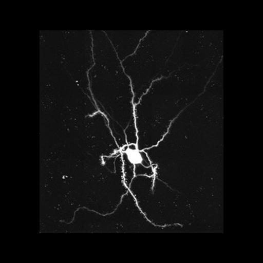
Tomographic reconstruction of a dendrite and associated dendritic spines from a medium spiny neuron from the mouse nucleus accumens. This image is a maximum intensity projection of a segment of dendrite in a 2 um section. The cell was filled by intracellular injection of Lucifer Yellow dye and photooxidized. This reconstructed image has been downsampled from the raw data image, which can be accessed using the link provided to the Cell Centered Database.

Technical Details

The tomogram was generated using an JEOL4000 IVEM. Single tilt images spanned -60 to 60° in 2° increments. Magnification, 3000X; accelerating voltage, 400KeV.

Biological Sources
NCBI Organism Classification
Mus musculus
Cell Type
CNS neuron (sensu Vertebrata)
Cellular Component
dendritic spine
dendrite
Attribution
Names
Maryann Martone
Eric Bushong
Link
CCDB 1
Citation
Digital Object Identifier (DOI)
doi:10.7295/W9CIL48401
Archival Resource Key (ARK)
ark:/b7295/w9cil48401
Grouping This image is part of a group.
Imaging
Image Type
recorded image
Image Mode
detection of electrons
illumination by electrons
Parameters Imaged
electron density
Data Qualifiers
processed data
Sample Preparation
Relation To Intact Cell
microtome-sectioned tissue
Dimensions
Spatial Axis Image Size Pixel Size
X 1440px 0.119µm
Y 1782px 0.119µm
Z 233px 0.5µm