Alternate header for print version


Licensing
Public Domain: This image is in the public domain and thus free of any copyright restrictions. However, as is the norm in scientific publishing and as a matter of courtesy, any user should credit the content provider for any public or private use of this image whenever possible. Learn more
tweet  
*CIL – Cell Image Library accession number. Please use this to reference an image.

CIL:44602*  Cite 
Description

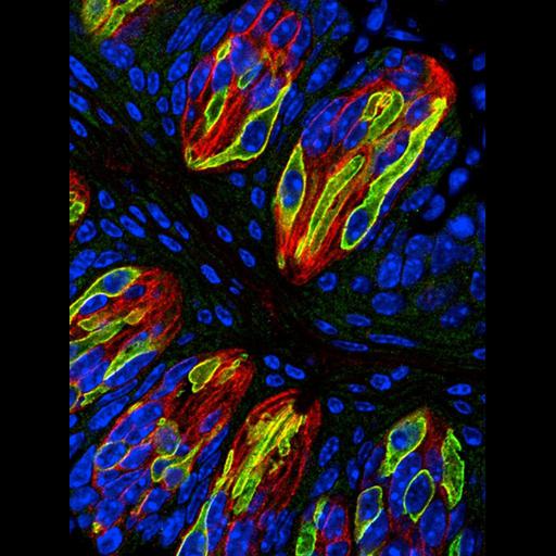
Fluorescence microscope image of a cryostat section of mouse tongue epithelium immuno-stained for all taste bud cells (red) type II taste bud cells (green)and counter-stained for DNA (blue). The image was selected for inclusion in the April 18th 2013 issue of the NIGMS Biomedical Beat that features noteworthy research results.

Technical Details

Anesthetized mice were perfused with 4% paraformaldehyde, tongue epithelium dissected out, frozen, and 8 micrometer cryostat sections cut. Sections were immuno-stained for all taste buds (red, anti-KCNQ1, Alexa 586) and type II taste buds (green, anti-TRPM5, Alexa 488), counter-stained with DAPI (blue), and recorded with a Zeiss LSM 710 confocal microscope equipped with 40x NA 1.1 water immersion objective lens. The image shows a single confocal plane with the red, green, and blue channels merged. See also: Akiyuki T et al. 2013. CALHM1 ion channel mediates purinergic neurotransmission of sweet, bitter and umami tastes. Nature 495:223-226.

Biological Sources
NCBI Organism Classification
Mus musculus
Cell Type
taste receptor cell
type II taste cell
Biological Context
Biological Process
olfaction
Attribution
Names
Kevin Foskett
Aki yuki Taruno
Link
NIGMS Biomedical Beat
Citation
Digital Object Identifier (DOI)
doi:10.7295/W9CIL44602
Archival Resource Key (ARK)
ark:/b7295/w9cil44602
Imaging
Image Type
recorded image
Image Mode
fluorescence microscopy
Parameters Imaged
fluorescence emission
Source of Contrast
distribution of epitope
Visualization Methods
Alexa Fluor 488
Alexa Fluor 568
4',6-diamidino-2-phenylindole (DAPI)
Data Qualifiers
processed data
suitable for spatial measurements
Sample Preparation
Methods
formaldehyde fixed tissue
Relation To Intact Cell
cryostat-sectioned tissue
Dimensions
Spatial Axis Image Size Pixel Size
X 650px 0.2µm
Y 868px 0.2µm