Alternate header for print version


Licensing
Attribution Non-Commercial; No Derivatives:This image is licensed under a Creative Commons Attribution, Non-Commercial, No Derivatives License. View License Deed | View Legal Code
tweet  
*CIL – Cell Image Library accession number. Please use this to reference an image.

CIL:41466*  Cite 
Description

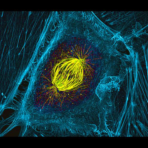
Confocal image of a mitotic spindle in a dividing cell. The spindle is shown in yellow and the surrounding actin cytoskeleton is in blue. Images were acquired using a 100X NA 1.4 phase objective lens and a Perkin Elmer spinning disc confocal scan head. A Hammamatsu Orca ER cooled CCD camera was used to collect images. Image collection was driven using Metamorph software. Complete Z-series of images were collected and deconvolved using AutoQuant software. The image is a maximum intensity projection of a subset of the entire Z-stack; the same subset of the Z-stack is used in each channel. Sixth Prize, 2007 Olympus BioScapes Digital Imaging Competition.

Technical Details

Biological Sources
NCBI Organism Classification
Sus scrofa domestica
Cell Type
epithelial cell
Cell Line
LLC-PK1
Cellular Component
spindle
actin cytoskeleton
Biological Context
Biological Process
mitosis
Attribution
Names
Patricia Wadsworth
2007 Olympus BioScapes Digital Imaging Competition
Link
2007 Olympus BioScapes Digital Imaging Compet...
OTHER
University of Massachusetts Amherst, MA, USA
Citation
Digital Object Identifier (DOI)
doi:10.7295/W9CIL41466
Archival Resource Key (ARK)
ark:/b7295/w9cil41466
Imaging
Image Type
recorded image
Image Mode
confocal microscopy
Parameters Imaged
fluorescence emission
Source of Contrast
distribution of epitope
Visualization Methods
phalloidin
primary antibody plus labeled secondary antibody
Fluorescein (FITC)
Rhodamine
Processing History
deconvolution
maximum projection
Sample Preparation
Methods
glutaraldehyde fixed tissue
Relation To Intact Cell
dispersed cells in vitro
Dimensions
Spatial Axis Image Size Pixel Size
X 1908px ——
Y 1620px ——