Alternate header for print version


Licensing
Attribution Non-Commercial Share Alike:This image is licensed under a Creative Commons Attribution, Non-Commercial Share Alike License. View License Deed | View Legal Code
tweet  
*CIL – Cell Image Library accession number. Please use this to reference an image.

CIL:40992*  Cite 
Description

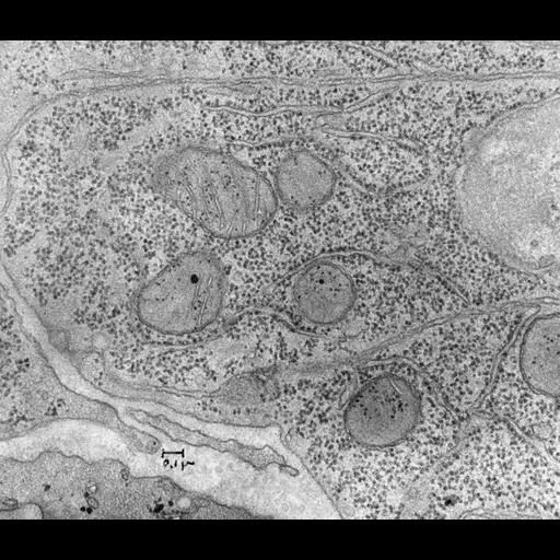
Transmission electron micrograph of a thin section of mouse intestine showing part of a cell with nucleus at left. The cytoplasm contains both membrane-associated polysomes (rough endoplasmic reticulum RER, sometimes termed granular endoplasmic reticulum) and free polysomes. Sen.

Technical Details

Original 3.25 in. x 4 in. lantern slides were scanned at 600dpi.

Biological Sources
NCBI Organism Classification
Mus musculus
Cell Type
intestinal cell
Cellular Component
rough endoplasmic reticulum
ribosome
nucleus
mitochondrion
plasma membrane
Biological Context
Biological Process
protein synthesis
cytoplasm organization
Attribution
Names
George E. Palade
Link
George E. Palade EM Slide Collection
Citation
Digital Object Identifier (DOI)
doi:10.7295/W9CIL40992
Archival Resource Key (ARK)
ark:/b7295/w9cil40992
Grouping This image is part of a group.
Imaging
Image Type
film
Image Mode
transmission electron microscopy (TEM)
Parameters Imaged
electron density
Source of Contrast
differences in adsorption or binding of stain
Visualization Methods
uranyl salt
osmium tetroxide
lead salt
Data Qualifiers
processed data
Sample Preparation
Methods
chemically fixed tissue
tissue in plastic embedment
Relation To Intact Cell
microtome-sectioned tissue
Dimensions
Spatial Axis Image Size Pixel Size
X 4470px ——
Y 3826px ——