Alternate header for print version


Licensing
This image is copyright protected. Any public or private use of this image is subject to prevailing copyright laws. Please contact the content provider of this image for permission requests.
tweet  
*CIL – Cell Image Library accession number. Please use this to reference an image.

CIL:40613*  Cite 
Description

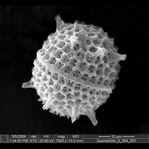
Scanning electron micrograph of a diatom frustule. The frustule is the hard and porous cell wall or external layer of diatoms.

Technical Details

Image collected on a FEI Quanta Family SEM with the following parameters: Magnification: 7000 x, Vacuum: 5.5 -5tprr, Voltage: 20 kv, Detector: ETD, and Working Distance: 15 mm.

Biological Sources
NCBI Organism Classification
Bacillariophyta
Cellular Component
frustule
Biological Context
Biological Process
frustule organization
Attribution
Names
Ekaterina Nikitina
Link
FEI Life Science Images
Citation
Digital Object Identifier (DOI)
doi:10.7295/W9CIL40613
Archival Resource Key (ARK)
ark:/b7295/w9cil40613
Imaging
Image Type
recorded image
Image Mode
scanning electron microscopy (SEM)
Parameters Imaged
elastic scattering of electrons
Processing History
unprocessed raw data
Sample Preparation
Relation To Intact Cell
whole mounted tissue
Dimensions
Spatial Axis Image Size Pixel Size
X 1024px ——
Y 943px ——