Alternate header for print version


Licensing
Attribution Non-Commercial Share Alike:This image is licensed under a Creative Commons Attribution, Non-Commercial Share Alike License. View License Deed | View Legal Code
tweet  
*CIL – Cell Image Library accession number. Please use this to reference an image.

CIL:40407*  Cite 
Description

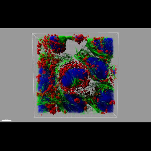
A confocal image of dividing keratinocyte cells labelled with mitotracker (red), tubulin (green) and nuclei (Dapi-blue). Cells were part of a study of the effect of Papilloma viruses in keratinocytes.

Biological Sources
Cell Type
keratinocyte
Cellular Component
mitochondrion
microtubule
nucleus
Biological Context
Biological Process
cytoskeleton organization
cell division
Attribution
Link
Andor Insight Awards
Andor Technology
Bitplane
OTHER
Kate Sullivan
NIMR
Citation
Digital Object Identifier (DOI)
doi:10.7295/W9CIL40407
Archival Resource Key (ARK)
ark:/b7295/w9cil40407
Imaging
Image Type
recorded image
Image Mode
confocal microscopy
Parameters Imaged
fluorescence emission
Source of Contrast
distribution of epitope
Visualization Methods
4',6-diamidino-2-phenylindole (DAPI)
probe for mitochondria
Processing History
projection
rendering
Sample Preparation
Relation To Intact Cell
dispersed cells in vitro
Dimensions
Spatial Axis Image Size Pixel Size
X 1652px ——
Y 1030px ——