Alternate header for print version


Licensing
Public Domain: This image is in the public domain and thus free of any copyright restrictions. However, as is the norm in scientific publishing and as a matter of courtesy, any user should credit the content provider for any public or private use of this image whenever possible. Learn more
tweet  
*CIL – Cell Image Library accession number. Please use this to reference an image.

CIL:40386*  Cite 
Description

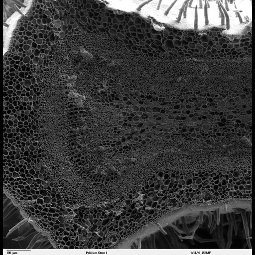
Scanning electron microscope image of cross-section through a Psidium guajava stem. The image shows the thin outer epidermal layer with a thick layer of cortex beneath. The vascular area consists of a secondary phloem and primary xylem, with vascular cambium in between. The central pith has some mucilage present. This image is part of a group on botanical stems (CIL:40378-40395).

Technical Details

Image collected on a Zeiss DSM 962 SEM. Complete specimen preparation protocol available at: http://remf.dartmouth.edu:8080/EM-Wiki/36

Biological Sources
NCBI Organism Classification
Psidium guajava
Cellular Component
mucilage
cortex
Biological Context
Biological Process
plant stem organization
Plant Growth
phloem
xylem
vascular cambium
Attribution
Names
Louisa Howard
Link
Dartmouth College EM Facility
Citation
Digital Object Identifier (DOI)
doi:10.7295/W9CIL40386
Archival Resource Key (ARK)
ark:/b7295/w9cil40386
Grouping This image is part of a group.
Sample Preparation
Methods
glutaraldehyde fixed tissue
ethanol dehydration
critical_point dried specimen
Relation To Intact Cell
sectioned tissue
Dimensions
Spatial Axis Image Size Pixel Size
X 1024px ——
Y 1049px ——