Alternate header for print version


Licensing
Public Domain: This image is in the public domain and thus free of any copyright restrictions. However, as is the norm in scientific publishing and as a matter of courtesy, any user should credit the content provider for any public or private use of this image whenever possible. Learn more
tweet  
*CIL – Cell Image Library accession number. Please use this to reference an image.

CIL:39781*  Cite 
Description

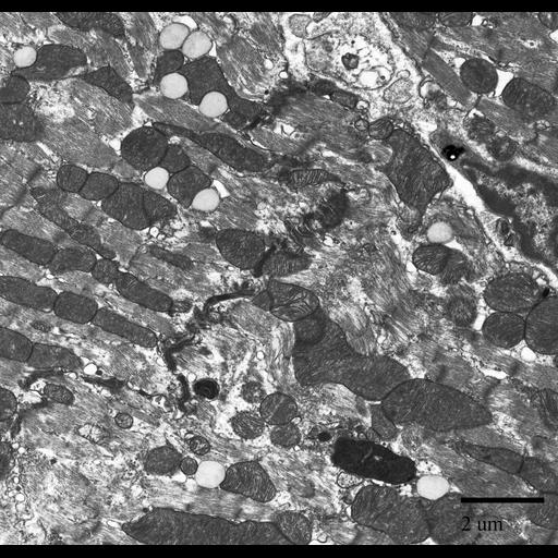
Transmission electron micrograph of ventricle tissue of a mouse cardiac muscle. This image shows the intercalated discs between the muscle fibers as well as many mitochondria and lipid droplets.

Technical Details

Primary fixation included: 2.5 % glutaraldehyde, 2% formaldehyde in 0.1 M Na-phosphate buffer, pH 7.4. Post-fixed in 2% OSO4 in 0.1 M Na-phosphate buffer, pH 7.4. Stained en bloc in 1% uranyl acetate. The tissue was then dehydrated in a graded series of ethanol and infiltrated with Spurr’s resin. Thin sections of 70 nm were trimmed using a diamond knife and post-stained in uranyl acetate and lead citrate. This micrograph image was taken using a Phillips CM 100 transmission electron microscope at an accelerating voltage of 80kV.

Biological Sources
NCBI Organism Classification
Mus musculus
Cell Type
myofibril
Cellular Component
I band
mitochondrion
lipid particle
Biological Context
Biological Process
muscle contraction
Mouse Gross Anatomy
TS25,ventricle
Attribution
Names
Dee Lauzon
Sue Lancelle
Marian Rice
OTHER
Mount Holyoke College
Citation
Digital Object Identifier (DOI)
doi:10.7295/W9CIL39781
Archival Resource Key (ARK)
ark:/b7295/w9cil39781
Grouping This image is part of a group.
Imaging
Image Type
recorded image
Image Mode
transmission electron microscopy (TEM)
Parameters Imaged
electron density
Source of Contrast
differences in adsorption or binding of stain
Visualization Methods
uranyl salt
lead salt
Processing History
unprocessed raw data
Sample Preparation
Methods
glutaraldehyde fixed tissue
formaldehyde fixed tissue
ethanol dehydration
tissue in epoxy resin embedment
Relation To Intact Cell
sectioned tissue
Dimensions
Spatial Axis Image Size Pixel Size
X 1770px ——
Y 1692px ——