Alternate header for print version


Licensing
Public Domain: This image is in the public domain and thus free of any copyright restrictions. However, as is the norm in scientific publishing and as a matter of courtesy, any user should credit the content provider for any public or private use of this image whenever possible. Learn more
tweet  
*CIL – Cell Image Library accession number. Please use this to reference an image.

CIL:39340*  Cite 
Description

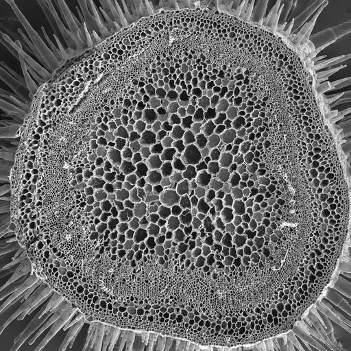
Scanning electron microscope image of Nicotiana alata stem cross section. Image shows outer epidermal layer, followed by the cortex and then large vascular bundles. The vascular bundles contain the phloem (nearest the cortex) and xylem.

Technical Details

See http://remf.dartmouth.edu:8080/EM-Wiki/36 for technical protocol

Biological Sources
NCBI Organism Classification
Nicotiana alata
Biological Context
Biological Process
plant stem organization
Plant Growth
epidermal layer
stem cortex
phloem
xylem
Attribution
Names
Louisa Howard
Link
Dartmouth College EM facility
Citation
Digital Object Identifier (DOI)
doi:10.7295/W9CIL39340
Archival Resource Key (ARK)
ark:/b7295/w9cil39340
Imaging
Image Type
recorded image
Image Mode
scanning electron microscopy (SEM)
Parameters Imaged
elastic scattering of electrons
Source of Contrast
differences in deposition of metal shadow
Visualization Methods
uranyl salt
shadowing and plating
Processing History
unprocessed raw data
Sample Preparation
Methods
glutaraldehyde fixed tissue
formaldehyde fixed tissue
Relation To Intact Cell
whole mounted tissue
Dimensions
Spatial Axis Image Size Pixel Size
X 4000px ——
Y 4000px ——