Alternate header for print version


Licensing
Attribution Non-Commercial; No Derivatives:This image is licensed under a Creative Commons Attribution, Non-Commercial, No Derivatives License. View License Deed | View Legal Code
tweet  
*CIL – Cell Image Library accession number. Please use this to reference an image.

CIL:39103*  Cite 
Description

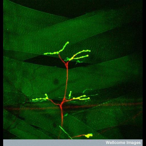
Confocal micrograph of an intact Drosophila larva imaged through the translucent cuticle showing the innervation of the dorsal (towards the back) muscle fibres by motor nerves. The muscles have been genetically engineered to express green fluorescent protein (GFP). The nerves express a red fluorescence protein (RFP), and the neuromuscular junctions are shown in yellow.

Technical Details

B0007255 Muscle Innervation, confocal micrograph 20/05/09. Wellcome Images available under the following creative commons usage http://creativecommons.org/licenses/by-nc/2.0/uk/

Biological Sources
NCBI Organism Classification
Drosophila
Cell Type
motor neuron
muscle cell
Cellular Component
neuromuscular junction
Biological Context
Biological Process
innervation
Attribution
Names
Hermann Aberle
Link
Wellcome Images
Citation
Digital Object Identifier (DOI)
doi:10.7295/W9CIL39103
Archival Resource Key (ARK)
ark:/b7295/w9cil39103
Imaging
Image Type
recorded image
Image Mode
confocal microscopy
Parameters Imaged
fluorescence emission
Source of Contrast
distribution of a specific protein
Visualization Methods
EGFP
mRFP1
Processing History
unprocessed raw data
Sample Preparation
Relation To Intact Cell
whole mounted tissue
Dimensions
Spatial Axis Image Size Pixel Size
X 550px ——
Y 576px ——