Alternate header for print version


Licensing
Attribution Non-Commercial; No Derivatives:This image is licensed under a Creative Commons Attribution, Non-Commercial, No Derivatives License. View License Deed | View Legal Code
tweet  
*CIL – Cell Image Library accession number. Please use this to reference an image.

CIL:39069*  Cite 
Description

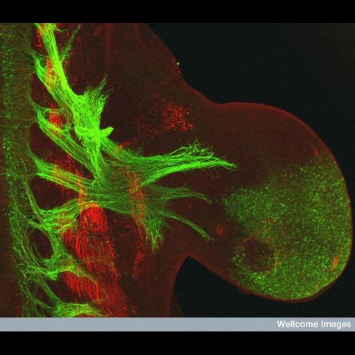
Confocal micrograph showing the development of motor neurons in the mouse forelimb at 11.5 days post coitum. The transgenic mice expresses a transgene of the Hb9 gene containing a genetic insert of the gene expressing green fluorescent protein (GFP). This transgenic insert allows the growth of motor neurons to be tracked. GFP is expressed where ever Hb9 is expressed and can be visualized using fluorescent immunohistochemistry techniques. This method can be used to accurately the time the motor neurons enter the limb to the point when they have branched to innervate all the developing muscles. In this image the developing motor neurons are shown in green (anti-GFP) and the muscle (myosin-32) is shown in red.

Technical Details

B0007552 Mouse forelimb motor neuron development 11.5 days post coitum. Wellcome Images available under the following creative commons usage http://creativecommons.org/licenses/by-nc-nd/2.0/uk/

Biological Sources
NCBI Organism Classification
Mus musculus
Cell Type
motor neuron
Cellular Component
neuron projection
myosin complex
Hb9 gene
myosin-32
Biological Context
Biological Process
neuron development
motor neuron growth
muscle structure development
Mouse Gross Anatomy
TS15,forelimb bud
Attribution
Link
Wellcome Images
OTHER
Clinical Sciences Centre, MRC
Citation
Digital Object Identifier (DOI)
doi:10.7295/W9CIL39069
Archival Resource Key (ARK)
ark:/b7295/w9cil39069
Imaging
Image Type
recorded image
Image Mode
confocal microscopy
Parameters Imaged
fluorescence emission
Source of Contrast
distribution of epitope
Visualization Methods
EGFP
primary antibody plus labeled secondary antibody
Processing History
unprocessed raw data
Sample Preparation
Methods
chemically fixed tissue
Relation To Intact Cell
whole mounted tissue
Dimensions
Spatial Axis Image Size Pixel Size
X 662px ——
Y 576px ——