Alternate header for print version


Licensing
Attribution Non-Commercial; No Derivatives:This image is licensed under a Creative Commons Attribution, Non-Commercial, No Derivatives License. View License Deed | View Legal Code
tweet  
*CIL – Cell Image Library accession number. Please use this to reference an image.

CIL:39043*  Cite 
Description

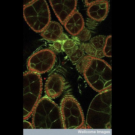
This fluorescent micrograph shows the developing egg chambers within the Drosophila melanogaster (fruit fly) ovary. The egg chambers house the maturing eggs - these are the cells at the tips of the egg chambers that face towards the outside of the image. The other cells present are called nurse cells; they occupy the rest of the chamber and assist the developing egg cells as they increase in size over time to produce a mature egg. The specimen has been stained with phalloidin, which marks the actin in the cells green and a nuclear marker that highlights the nuclei in red.

Technical Details

B0007685 Developing egg chambers from the Drosophila ovary. Wellcome Images available under the following creative commons usage http://creativecommons.org/licenses/by-nc-nd/2.0/uk/

Biological Sources
NCBI Organism Classification
Drosophila melanogaster
Cell Type
egg
invertebrate nurse cell
Cellular Component
actin cytoskeleton
nucleus
Attribution
Link
Wellcome Images
OTHER
National Institute for Medical Research, MRC
Citation
Digital Object Identifier (DOI)
doi:10.7295/W9CIL39043
Archival Resource Key (ARK)
ark:/b7295/w9cil39043
Imaging
Image Type
recorded image
Image Mode
fluorescence microscopy
Parameters Imaged
fluorescence emission
Source of Contrast
distribution of epitope
Visualization Methods
phalloidin
Processing History
unprocessed raw data
Sample Preparation
Methods
chemically fixed tissue
permeabilized tissue
Relation To Intact Cell
whole mounted tissue
Dimensions
Spatial Axis Image Size Pixel Size
X 379px ——
Y 576px ——