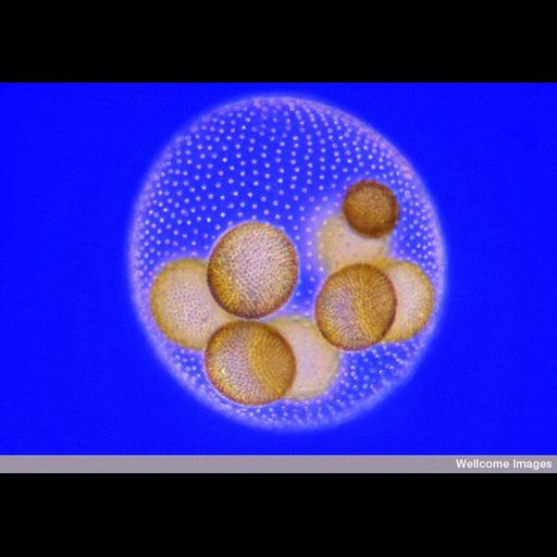
Light micrograph of Volvox taken using Rheinberg illumination. Volvox form large (approximately 500 µm) spherical colonies that can contain up to 15,000 biflagellate, photosynthetic cells embedded in their gelatinous walls. The colonies are hollow (coenobia) and often found with daughter colonies, and/or groups of gametes lying inside them. Beating of the flagella is strongly co-coordinated so that, for instance, colonies can rapidly react to a light source by swimming toward it. They are mainly found in freshwater ponds. The image is of a live specimen captured with a 0.5 microsecond flash.
B0007761 Colony of volvox with eight daughter coenobia. Wellcome Images available under the following creative commons usage http://creativecommons.org/licenses/by-nc-nd/2.0/uk/ See the following website for a description of Rheinberg illumination http://micro.magnet.fsu.edu/primer/techniques/rheinberg.html
| Spatial Axis | Image Size | Pixel Size |
|---|---|---|
| X | 800px | —— |
| Y | 561px | —— |