Alternate header for print version


Licensing
Attribution Non-Commercial; No Derivatives:This image is licensed under a Creative Commons Attribution, Non-Commercial, No Derivatives License. View License Deed | View Legal Code
tweet  
*CIL – Cell Image Library accession number. Please use this to reference an image.

CIL:39021*  Cite 
Description

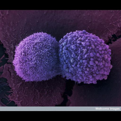
Colorized scanning electron micrograph showing two lung cancer cells. These cells were grown using cell culture techniques.

Technical Details

B0007782 Lung cancer cells. Wellcome Images available under the following creative commons usage http://creativecommons.org/licenses/by-nc-nd/2.0/uk/

Biological Sources
Cell Type
lung cancer cell
Cellular Component
cell surface
ruffle membrane
Biological Context
Biological Process
cell-cell adhesion
Human Disease
lung carcinoma
Attribution
Names
Anne Weston
Link
Wellcome Images
OTHER
London Research Institute, CRUK
Citation
Digital Object Identifier (DOI)
doi:10.7295/W9CIL39021
Archival Resource Key (ARK)
ark:/b7295/w9cil39021
Imaging
Image Type
recorded image
Image Mode
scanning electron microscopy (SEM)
Parameters Imaged
elastic scattering of electrons
Source of Contrast
differences in deposition of metal shadow
Visualization Methods
visualization of contiguous regions
Processing History
colorized
Sample Preparation
Methods
chemically fixed tissue
Relation To Intact Cell
dispersed cells in vitro
Dimensions
Spatial Axis Image Size Pixel Size
X 688px ——
Y 576px ——