Alternate header for print version


Licensing
Attribution Non-Commercial; No Derivatives:This image is licensed under a Creative Commons Attribution, Non-Commercial, No Derivatives License. View License Deed | View Legal Code
tweet  
*CIL – Cell Image Library accession number. Please use this to reference an image.

CIL:38980*  Cite 
Description

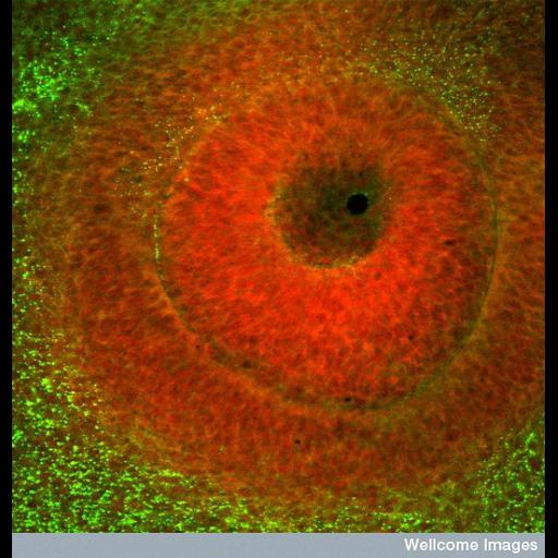
Confocal image of the developing eye of a chick embryo. The dark region in the centre is the where the surface layer has invaginated to form the lens vesicle. The tissue surrounding this space is stained red and will go on to form the lens. The outer ring of red stained cells will form the retina.

Technical Details

B0002683 Developing eye in a chick embryo. Wellcome Images available under the following creative commons usage http://creativecommons.org/licenses/by-nc-nd/2.0/uk/

Biological Sources
NCBI Organism Classification
Gallus gallus
Cell Type
retinal progenitor cell
lens epithelial cell
Cellular Component
cell surface
Biological Context
Biological Process
eye development
eye morphogenesis
Attribution
Names
Kate Whitley
Link
Wellcome Images
Citation
Digital Object Identifier (DOI)
doi:10.7295/W9CIL38980
Archival Resource Key (ARK)
ark:/b7295/w9cil38980
Imaging
Image Type
recorded image
Image Mode
confocal microscopy
Parameters Imaged
fluorescence emission
Source of Contrast
distribution of epitope
Processing History
unprocessed raw data
Sample Preparation
Relation To Intact Cell
whole mounted tissue
Dimensions
Spatial Axis Image Size Pixel Size
X 550px ——
Y 576px ——