Alternate header for print version


Licensing
Attribution Non-Commercial; No Derivatives:This image is licensed under a Creative Commons Attribution, Non-Commercial, No Derivatives License. View License Deed | View Legal Code
tweet  
*CIL – Cell Image Library accession number. Please use this to reference an image.

CIL:38930*  Cite 
Description

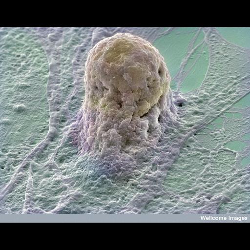
Colorized scanning electron micrograph of a human embryonic stem cell (gold) growing on a layer of supporting cells (fibroblasts). Stem cells are derived from very early embryos and can either be grown to stay in their original state or triggered to form almost any type of human cell. The fibroblasts provide special factors that maintain the stem cells in their original state. The stem cell appears to be grasped by the underlying fibroblast.

Technical Details

B0006219 Human embryonic stem cell. Wellcome Images available under the following creative commons usage http://creativecommons.org/licenses/by-nc-nd/2.0/uk/

Biological Sources
NCBI Organism Classification
Homo sapiens
Cell Type
embryonic stem cell
fibroblast
Cellular Component
cell surface
Attribution
Names
Annie Cavanagh
Link
Wellcome Images
Citation
Digital Object Identifier (DOI)
doi:10.7295/W9CIL38930
Archival Resource Key (ARK)
ark:/b7295/w9cil38930
Imaging
Image Type
recorded image
Image Mode
scanning electron microscopy (SEM)
Parameters Imaged
elastic scattering of electrons
Source of Contrast
differences in deposition of metal shadow
Visualization Methods
visualization of contiguous regions
Processing History
colorized
Sample Preparation
Methods
chemically fixed tissue
Relation To Intact Cell
dispersed cells in vitro
Dimensions
Spatial Axis Image Size Pixel Size
X 734px ——
Y 576px ——