Alternate header for print version


Licensing
Attribution Non-Commercial Share Alike:This image is licensed under a Creative Commons Attribution, Non-Commercial Share Alike License. View License Deed | View Legal Code
tweet  
*CIL – Cell Image Library accession number. Please use this to reference an image.

CIL:38599*  Cite 
Description

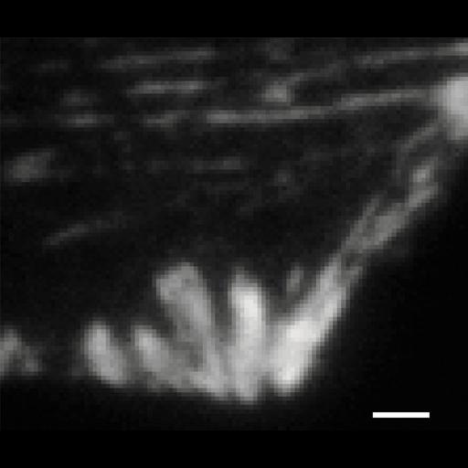
This total internal reflection (TIRF) image of both the Dronpa-alphaa ctinin and the unconverted tdEos-vinculin channels corresponds to the same image field as the diffraction limited DIC image CIL 38597 and the 2-color photoactivation localization microscopy (PALM) image 38598. Bar is 2 microns. Image made available by Catherine and James Galbraith and corresponds to Figure 2 in PNAS U S A. 2007 Dec 18;104(51):20308-13.

Biological Sources
NCBI Organism Classification
Homo sapiens
Cell Type
fibroblast
Cell Line
foreskin fibroblast
Cellular Component
focal adhesion
actin cytoskeleton
alpha actinin
vinculin
Biological Context
Biological Process
focal adhesion assembly
Attribution
Names
Hari Shroff
Catherine G Galbraith
James A. Galbraith
Helen White
Jennifer Gillette
Scott Olenych
Michael W. Davidson
Eric Betzig
Published
Proc Natl Acad Sci U S A. 2007 Dec 18;104(51):20308-13. Epub 2007 Dec 12. Erratum in: Proc Natl Acad Sci U S A. 2008 Sep 30;105(39):15220.
Pubmed
18077327
Link
article
Citation
Digital Object Identifier (DOI)
doi:10.7295/W9CIL38599
Archival Resource Key (ARK)
ark:/b7295/w9cil38599
Grouping This image is part of a group.
Imaging
Image Type
recorded image
Image Mode
TIRF (total internal reflection)
Parameters Imaged
fluorescence emission
Source of Contrast
distribution of a specific protein
Visualization Methods
Dronpa
tdEos
Processing History
unprocessed raw data
Data Qualifiers
raw, unprocessed data
Sample Preparation
Methods
formaldehyde fixed tissue
Relation To Intact Cell
dispersed cells in vitro
Dimensions
Spatial Axis Image Size Pixel Size
X 894px ——
Y 714px ——