Alternate header for print version


Licensing
Attribution Non-Commercial Share Alike:This image is licensed under a Creative Commons Attribution, Non-Commercial Share Alike License. View License Deed | View Legal Code
tweet  
*CIL – Cell Image Library accession number. Please use this to reference an image.

CIL:37298*  Cite 
Description

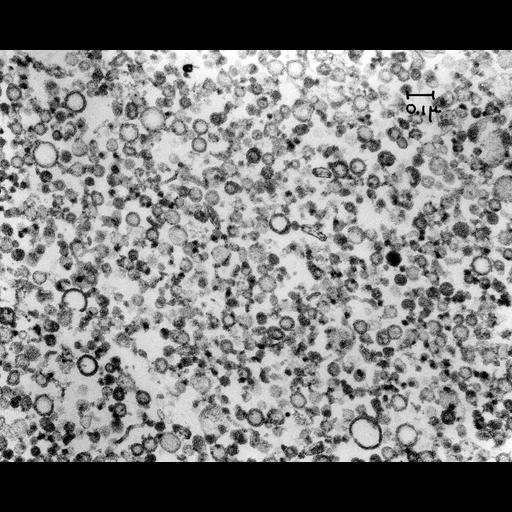
Transmission electron micrograph of smooth microsomes obtained by fraction of guinea pig pancreas. Cell fractionation confirmed that newly synthesized acinar cell secretory proteins moved from the rough endoplasmic reticulum to the periphery of the Golgi in the lumen of a smooth-surfaced membrane bounded compartment. Image made available by James D. Jamieson and the Department of Cell Biology, Yale University School of Medicine.

Technical Details

Additional technical details regarding cell fractionation, specimen processing, and electron microscopy can be found in: Jamieson, J.D., and Palade, G.E. J. Cell Biol. 34:577-596, 1967. Original 3.25 in. x 4 in. lantern slides were scanned at 600dpi. Original Magnification: x10,000

Biological Sources
NCBI Organism Classification
Cavia porcellus
Cell Type
pancreatic cell
Cellular Component
smooth microsome
Biological Context
Biological Process
protein secretion
Attribution
Names
James D. Jamieson
Link
George E. Palade EM Slide COllection
Citation
Digital Object Identifier (DOI)
doi:10.7295/W9CIL37298
Archival Resource Key (ARK)
ark:/b7295/w9cil37298
Grouping This image is part of a group.
Imaging
Image Type
recorded image
Image Mode
transmission electron microscopy (TEM)
Parameters Imaged
electron density
Source of Contrast
differences in adsorption or binding of stain
Visualization Methods
lead salt
uranyl salt
Processing History
scanned negative
Sample Preparation
Methods
osmium tetroxide fixed tissue
Relation To Intact Cell
isolated subcellular component
Dimensions
Spatial Axis Image Size Pixel Size
X 6000px ——
Y 4834px ——