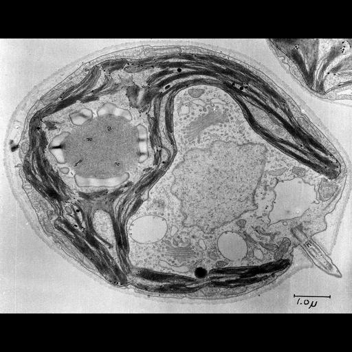
An electron micrograph showing a central longitudinal section of the unicellular green alga Chlamydomonas reinhardtii (y-1 yellow mutant) grown in the light. The single cup shaped chloroplast with pyrenoid (storage granule) surrounds the central nucleus and other organelles. Osmiophilic globules and a few starch granules are scattered among the thylakoid membranes. The pyrenoid appears as a large, finely granular mass of polygonal profile surrounded by discontinuous shell of starch plates and penetrated by a system of tubules. Surrounded by the chloroplast are the nucleus, dictyosomes (Golgi bodies) and their associated vacuoles, and endoplasmic reticulum cisternae of transitional type. Mitochondria are concentrated at the anterior pole of the cell and between the chloroplast and the cell membrane. A contractile vacuole and a flagellum are also visible.
Image corresponds to figure 2 in J Cell Biol. 1967 Dec;35(3):521-52. Image made available by James D. Jamieson and the Department of Cell Biology, Yale University School of Medicine. Original 3.25 in. x 4 in. lantern slides were scanned at 600dpi. Original magnification 8,400.
| Spatial Axis | Image Size | Pixel Size |
|---|---|---|
| X | 6000px | —— |
| Y | 4694px | —— |