Alternate header for print version


Licensing
Attribution Non-Commercial Share Alike:This image is licensed under a Creative Commons Attribution, Non-Commercial Share Alike License. View License Deed | View Legal Code
tweet  
*CIL – Cell Image Library accession number. Please use this to reference an image.

CIL:37168*  Cite 
Description

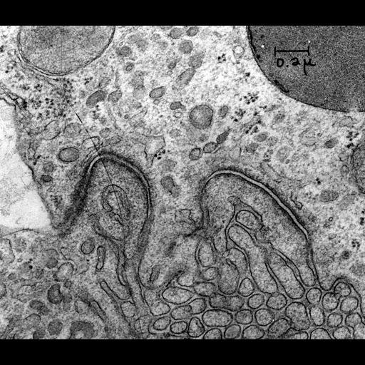
Transmission electron micrograph of adherens junctions (macula adherens or desmosome)in the proximal tubule of a rat kidney. The junctions with associated tonofilaments are seen at the apposed plasma membranes between two epithelial cells. Image made available by James D. Jamieson and the Department of Cell Biology, Yale University School of Medicine.

Technical Details

Original 3.25 in. x 4 in. lantern slides were scanned at 600dpi. Original Magnification: x35,000.

Biological Sources
NCBI Organism Classification
Rattus
Cell Type
kidney tubule cell
Cellular Component
adherens junction
desmosome
Biological Context
Biological Process
adherens junction organization
Attribution
Names
Marilyn G. Farquhar
Link
George E. Palade EM Slide Collection
Citation
Digital Object Identifier (DOI)
doi:10.7295/W9CIL37168
Archival Resource Key (ARK)
ark:/b7295/w9cil37168
Grouping This image is part of a group.
Imaging
Image Type
recorded image
Image Mode
transmission electron microscopy (TEM)
Parameters Imaged
electron density
Source of Contrast
differences in adsorption or binding of stain
Visualization Methods
osmium tetroxide
Processing History
unprocessed raw data
Data Qualifiers
raw, unprocessed data
Sample Preparation
Methods
chemically fixed tissue
Relation To Intact Cell
sectioned tissue
Dimensions
Spatial Axis Image Size Pixel Size
X 6000px ——
Y 5166px ——