Alternate header for print version


Licensing
Attribution Non-Commercial Share Alike:This image is licensed under a Creative Commons Attribution, Non-Commercial Share Alike License. View License Deed | View Legal Code
tweet  
*CIL – Cell Image Library accession number. Please use this to reference an image.

CIL:37120*  Cite 
Description

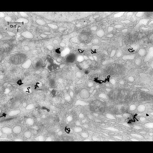
Electron microscope autoradiograph taken 20 min after injection showing that the radiographic label has migrated from the rough endoplasmic reticulum to the Golgi complex. Most of the radioactive grains are over condensing vacuoles and the perpheral clusters of small vesicles have much less activity. Image corresponds to Figure 5 in J. Cell Biol. 20:473-495. Image made available by James D. Jamieson and the Department of Cell Biology, Yale University School of Medicine.

Technical Details

Anesthetized animals were intravenously injected with 1-5 millicuris of DL-leucine 4,5-H^3 with a specific activity of 3570 mc/mM. At times ranging from 4 min to 15 hrs after injection the pancreas was removed and fixed in osmium tetroxide. Tissue was embedded in methacrylate, sectioned, and post-stained with uranyl acetate. Original 3.25 in. x 4 in. lantern slides were scanned at 600dpi. Original Magnification: x30,000.

Biological Sources
NCBI Organism Classification
Cavia porcellus
Cell Type
pancreas cell
Cellular Component
mitochondrion
Golgi-associated vesicle
Biological Context
Biological Process
protein secretion
protein transport
Attribution
Names
Lucien Caro
Published
J Cell Biol. 1964 Mar;20:473-95.
Pubmed
14128049
Link
George E. Palade EM Slide Collection
Citation
Digital Object Identifier (DOI)
doi:10.7295/W9CIL37120
Archival Resource Key (ARK)
ark:/b7295/w9cil37120
Grouping This image is part of a group.
Imaging
Image Type
recorded image
Image Mode
transmission electron microscopy (TEM)
electron microscope autoradiograph
Parameters Imaged
electron density
Source of Contrast
differences in adsorption or binding of stain
Visualization Methods
uranyl salt
metabolically incorporated radioisotope
Processing History
unprocessed raw data
Sample Preparation
Methods
osmium tetroxide fixed tissue
Relation To Intact Cell
sectioned tissue
Dimensions
Spatial Axis Image Size Pixel Size
X 6000px ——
Y 5096px ——