Alternate header for print version


Licensing
Public Domain: This image is in the public domain and thus free of any copyright restrictions. However, as is the norm in scientific publishing and as a matter of courtesy, any user should credit the content provider for any public or private use of this image whenever possible. Learn more
tweet  
*CIL – Cell Image Library accession number. Please use this to reference an image.

CIL:36254*  Cite 
Description

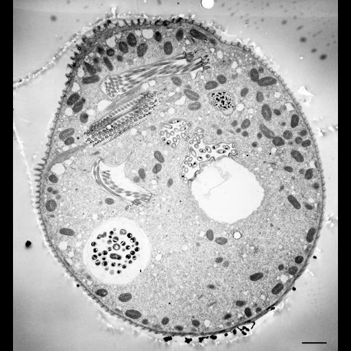
The 11 micrographs of the group are views of a serially-sectioned contracted Vorticella convallaria cell that show the main features of this cell. This figure shows 1 food vacuole; micronucleus; contractile vacuole connected by 2 CV pores to the bacteria-filled chamber; peristome; perikinetids of the perioral cilia. TEM taken on 4/1/71 by R. Allen with Hitachi HU11A operating at 75kV. Neg. 3,400X. Bar = 2µm. The negative was printed to paper and the image was scanned to Photoshop. This image is suitable for qualitative analysis. A high resolution version of this image in the library (CIL:40570) is available for quantitative analysis. Additional information available at (http://www5.pbrc.hawaii.edu/allen/).

Technical Details

Standard glutaraldehyde fixation followed by osmium tetroxide, dehydrated in alcohol and embedded in an epoxy resin. Microtome sections prepared at approximately 75nm thickness. Additional information available at (http://www5.pbrc.hawaii.edu/allen/).

Biological Sources
NCBI Organism Classification
Vorticella convallaria
Cell Type
cell by organism
eukaryotic cell
Eukaryotic Protist
Ciliated Protist
Cell Line
Cambridge Culture Collection, Cambridge, England, # LB1690/1
Cellular Component
cell
food vacuole
micronucleus
contractile vacuole
contractile vacuole pore
oral apparatus
food vacuole
Attribution
Names
Richard Allen (University of Hawaii)
Citation
Digital Object Identifier (DOI)
doi:10.7295/W9CIL36254
Archival Resource Key (ARK)
ark:/b7295/w9cil36254
Grouping This image is part of a group.
Imaging
Image Type
transmission electron microscopy (TEM)
illumination by electrons
Image Mode
detection of electrons
film
Parameters Imaged
electron density
Source of Contrast
stain with broad specificity
Visualization Methods
stain with broad specificity
osmium tetroxide
uranyl salt
lead salt
Processing History
recorded image
film
Print from negative scanned for Photoshop.
Data Qualifiers
processed data
Dimensions
Spatial Axis Image Size Pixel Size
X 2737px ——
Y 2952px ——