Alternate header for print version


Licensing
Attribution Non-Commercial; No Derivatives:This image is licensed under a Creative Commons Attribution, Non-Commercial, No Derivatives License. View License Deed | View Legal Code
tweet  
*CIL – Cell Image Library accession number. Please use this to reference an image.

CIL:36065*  Cite 
Description

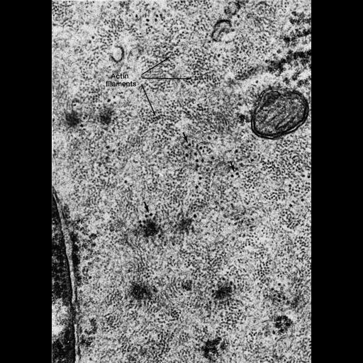
Figure 433 from Chapter 16 (Cytoplasmic matrix and cytoskeleton) of 'The Cell, 2nd Ed.' by Don W. Fawcett M.D. Smooth muscle of mouse epididymal duct. This micrograph is a high magnification view of actin filaments in cross section from part of the accompanying image CIL:36064 in this image group. In contrast to striated muscle, actin filaments in smooth muscle occur in irregular groups that represent bundles or tracts of parallel flaments. A PDF copy of the accompanying chapter is available on the ASCB’s BioEDUCATE website.

Biological Sources
NCBI Organism Classification
Mus musculus
Cell Type
smooth muscle cell
Cellular Component
actin filament
cytoplasm
ribosome
Biological Context
Biological Process
smooth muscle contraction
Attribution
Names
Don W. Fawcett
Published
ISBN 0721635849
Link
http://www.ascb.org/bioeducate/FawcettTheCell...
Citation
Digital Object Identifier (DOI)
doi:10.7295/W9CIL36065
Archival Resource Key (ARK)
ark:/b7295/w9cil36065
Grouping This image is part of a group.
Imaging
Image Type
transmission electron microscopy (TEM)
Image Mode
illumination by electrons
detection of electrons
Parameters Imaged
electron density
Source of Contrast
stain with broad specificity
Visualization Methods
osmium tetroxide
Data Qualifiers
processed data
Dimensions
Spatial Axis Image Size Pixel Size
X 892px ——
Y 1248px ——