Alternate header for print version


Licensing
Attribution Non-Commercial; No Derivatives:This image is licensed under a Creative Commons Attribution, Non-Commercial, No Derivatives License. View License Deed | View Legal Code
tweet  
*CIL – Cell Image Library accession number. Please use this to reference an image.

CIL:36045*  Cite 
Description

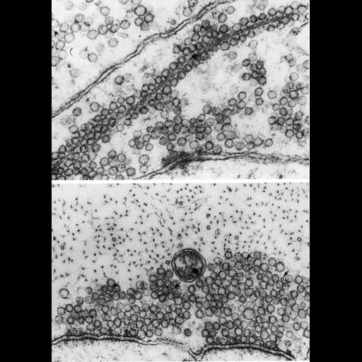
Figures 408 (upper) and 409 (lower) from Chapter 16 (Cytoplasmic matrix and cytoskeleton) of 'The Cell, 2nd Ed.' by Don W. Fawcett M.D. Upper panel: Vesicles, presumably in transport along cytoplasmic microtubules in a giant axon from the central nervous system of the lamprey. Lower panel: vesicles are also clustered around microtubules (arrows) at the periphery of sites of synaptic contact. Figures from Smith, Järlfors, Beránek, J. Cell Biol. 46:199-219, 1970 (PMID:5452412). A PDF copy of the accompanying chapter is available on the ASCB’s BioEDUCATE website.

Biological Sources
NCBI Organism Classification
Petromyzon marinus
Cell Type
Mauthner neuron
Cellular Component
microtubule
giant axon
transport vesicle
Biological Context
Biological Process
microtubule-based transport
Attribution
Names
Don W. Fawcett
D.S. Smith
U. Järlfors
R. Beránek
Published
ISBN 0721635849
Link
http://www.ascb.org/bioeducate/FawcettTheCell...
Citation
Digital Object Identifier (DOI)
doi:10.7295/W9CIL36045
Archival Resource Key (ARK)
ark:/b7295/w9cil36045
Grouping This image is part of a group.
Imaging
Image Type
transmission electron microscopy (TEM)
Image Mode
illumination by electrons
detection of electrons
Parameters Imaged
electron density
Source of Contrast
stain with broad specificity
Visualization Methods
electron-dense stain
Data Qualifiers
processed data
Dimensions
Spatial Axis Image Size Pixel Size
X 892px ——
Y 1252px ——