Alternate header for print version


Licensing
Attribution Non-Commercial; No Derivatives:This image is licensed under a Creative Commons Attribution, Non-Commercial, No Derivatives License. View License Deed | View Legal Code
tweet  
*CIL – Cell Image Library accession number. Please use this to reference an image.

CIL:36015*  Cite 
Description

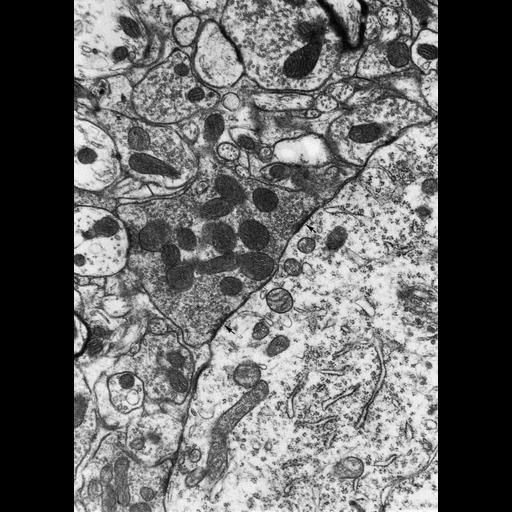
Figure 397 from Chapter 15 (Cytoplasmic Inclusions) of 'The Cell, 2nd Ed.' by Don W. Fawcett M.D. Axosomatic and axodendritic synapses in the central nervous system of the rat. A nerve terminal in the center of the micrograph can be seen making contact with a cell body (occupying the lower right; arrows indicate two regions of apparent active zone contact. Notably, there is also synaptic contact by same axon with a dendrite to the left. A second axodendritic synapse is present at the top of the micrograph. Image by Enrico Mugnaini. A PDF copy of the accompanying chapter is available on the ASCB’s BioEDUCATE website.

Biological Sources
NCBI Organism Classification
Rattus
Cell Type
CNS neuron (sensu Vertebrata)
Cellular Component
synapse
axon terminus
neuronal cell body
synaptic vesicle
mitochondrion
Biological Context
Biological Process
synapse organization
Attribution
Names
Don W. Fawcett
Enrico Mugnaini
Published
ISBN 0721635849
Link
http://www.ascb.org/bioeducate/FawcettTheCell...
Citation
Digital Object Identifier (DOI)
doi:10.7295/W9CIL36015
Archival Resource Key (ARK)
ark:/b7295/w9cil36015
Grouping This image is part of a group.
Imaging
Image Type
transmission electron microscopy (TEM)
Image Mode
illumination by electrons
detection of electrons
Parameters Imaged
electron density
Source of Contrast
stain with broad specificity
Visualization Methods
osmium tetroxide
Data Qualifiers
processed data
Dimensions
Spatial Axis Image Size Pixel Size
X 889px ——
Y 1249px ——