Alternate header for print version


Licensing
Attribution Non-Commercial; No Derivatives:This image is licensed under a Creative Commons Attribution, Non-Commercial, No Derivatives License. View License Deed | View Legal Code
tweet  
*CIL – Cell Image Library accession number. Please use this to reference an image.

CIL:36005*  Cite 
Description

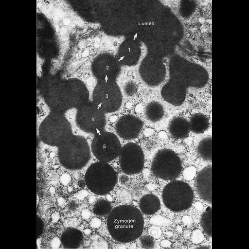
Figure 387 from Chapter 15 (Cytoplasmic Inclusions) of 'The Cell, 2nd Ed.' by Don W. Fawcett M.D. The apical region of an acinar cell from perfused canine pancreas stimulated with pancreozymin shows a chain of vesicles on a secretory path to the surface of the cell. Image from A. Ichikawa, J. Cell Biol. 24: 369, 1965 (PMID:14326122). A PDF copy of the accompanying chapter is available on the ASCB’s BioEDUCATE website.

Biological Sources
NCBI Organism Classification
Canis lupus familiaris
Cell Type
acinar cell
simple columnar epithelial cell
Cellular Component
secretory granule
Biological Context
Biological Process
exocytosis
secretory granule organization
Attribution
Names
Don W. Fawcett
A. Ichikawa
Published
ISBN 0721635849
Link
http://www.ascb.org/bioeducate/FawcettTheCell...
Citation
Digital Object Identifier (DOI)
doi:10.7295/W9CIL36005
Archival Resource Key (ARK)
ark:/b7295/w9cil36005
Grouping This image is part of a group.
Imaging
Image Type
transmission electron microscopy (TEM)
Image Mode
illumination by electrons
detection of electrons
Parameters Imaged
electron density
Source of Contrast
stain with broad specificity
Visualization Methods
osmium tetroxide
Data Qualifiers
processed data
Dimensions
Spatial Axis Image Size Pixel Size
X 894px ——
Y 1266px ——