Alternate header for print version


Licensing
Attribution Non-Commercial; No Derivatives:This image is licensed under a Creative Commons Attribution, Non-Commercial, No Derivatives License. View License Deed | View Legal Code
tweet  
*CIL – Cell Image Library accession number. Please use this to reference an image.

CIL:35997*  Cite 
Description

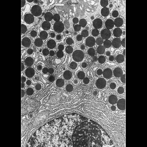
Figure 376 from Chapter 15 (Cytoplasmic Inclusions) of 'The Cell, 2nd Ed.' by Don W. Fawcett M.D. This human pancreatic acinar cell has zymogen granules crowded toward the apex. The endoplasmic reticulum cisternae that are interspersed between these secretory vesicles are more variable in width, and less highly ordered than those at the cell base. Image by Susumo Ito; Arthur Like. A PDF copy of the accompanying chapter is available on the ASCB’s BioEDUCATE website.

Biological Sources
NCBI Organism Classification
Homo sapiens
Cell Type
acinar cell
Cellular Component
secretory granule
zymogen granule
endoplasmic reticulum
Biological Context
Biological Process
secretory granule organization
Attribution
Names
Don W. Fawcett
Susumo Ito; Arthur Like
Published
ISBN 0721635849
Link
http://www.ascb.org/bioeducate/FawcettTheCell...
Citation
Digital Object Identifier (DOI)
doi:10.7295/W9CIL35997
Archival Resource Key (ARK)
ark:/b7295/w9cil35997
Grouping This image is part of a group.
Imaging
Image Type
transmission electron microscopy (TEM)
Image Mode
illumination by electrons
detection of electrons
Parameters Imaged
electron density
Source of Contrast
stain with broad specificity
Visualization Methods
osmium tetroxide
Data Qualifiers
processed data
Dimensions
Spatial Axis Image Size Pixel Size
X 897px ——
Y 1257px ——