Alternate header for print version


Licensing
Attribution Non-Commercial; No Derivatives:This image is licensed under a Creative Commons Attribution, Non-Commercial, No Derivatives License. View License Deed | View Legal Code
tweet  
*CIL – Cell Image Library accession number. Please use this to reference an image.

CIL:35990*  Cite 
Description

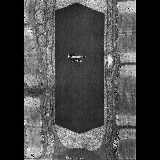
Figure 369 from Chapter 15 (Cytoplasmic Inclusions) of 'The Cell, 2nd Ed.' by Don W. Fawcett M.D. Erythrocytes in a longitudinal section through a capillary from the myocardium of a cat. In the upper cell, the hemoglobin has formed a large single crystal, and the surrounding cytoplasm is pale. In the lower erythrocyte, hemoglobin appears to be normally distributed, and the cytoplasmic matrix appears uniform. A PDF copy of the accompanying chapter is available on the ASCB’s BioEDUCATE website.

Biological Sources
NCBI Organism Classification
Felis catus
Cell Type
erythrocyte
Cellular Component
inclusion body
Biological Context
Biological Process
hemoglobin catabolic process
Attribution
Names
Don W. Fawcett
Published
ISBN 0721635849
Link
http://www.ascb.org/bioeducate/FawcettTheCell...
Citation
Digital Object Identifier (DOI)
doi:10.7295/W9CIL35990
Archival Resource Key (ARK)
ark:/b7295/w9cil35990
Grouping This image is part of a group.
Imaging
Image Type
transmission electron microscopy (TEM)
Image Mode
illumination by electrons
detection of electrons
Parameters Imaged
electron density
Source of Contrast
stain with broad specificity
Visualization Methods
osmium tetroxide
Data Qualifiers
processed data
Dimensions
Spatial Axis Image Size Pixel Size
X 888px ——
Y 1267px ——