Alternate header for print version


Licensing
Attribution Non-Commercial; No Derivatives:This image is licensed under a Creative Commons Attribution, Non-Commercial, No Derivatives License. View License Deed | View Legal Code
tweet  
*CIL – Cell Image Library accession number. Please use this to reference an image.

CIL:35978*  Cite 
Description

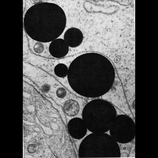
Figure 356 from Chapter 15 (Cytoplasmic Inclusions) of 'The Cell, 2nd Ed.' by Don W. Fawcett M.D. Lipid appears as spherical droplets in the cytosol, shown here in a Sertoli cell from the oppossum testis. In this preparation, staining of the lipid was intensified with osmium. A PDF copy of the accompanying chapter is available on the ASCB’s BioEDUCATE website.

Biological Sources
NCBI Organism Classification
Didelphis virginiana
Cell Type
Sertoli cell
Cellular Component
lipid particle
Biological Context
Biological Process
lipid storage
Attribution
Names
Don W. Fawcett
Published
ISBN 0721635849
Link
http://www.ascb.org/bioeducate/FawcettTheCell...
Citation
Digital Object Identifier (DOI)
doi:10.7295/W9CIL35978
Archival Resource Key (ARK)
ark:/b7295/w9cil35978
Grouping This image is part of a group.
Imaging
Image Type
transmission electron microscopy (TEM)
Image Mode
illumination by electrons
detection of electrons
Parameters Imaged
electron density
Source of Contrast
stain with broad specificity
Visualization Methods
osmium tetroxide
Data Qualifiers
processed data
Dimensions
Spatial Axis Image Size Pixel Size
X 897px ——
Y 1261px ——