Alternate header for print version


Licensing
Attribution Non-Commercial; No Derivatives:This image is licensed under a Creative Commons Attribution, Non-Commercial, No Derivatives License. View License Deed | View Legal Code
tweet  
*CIL – Cell Image Library accession number. Please use this to reference an image.

CIL:35972*  Cite 
Description

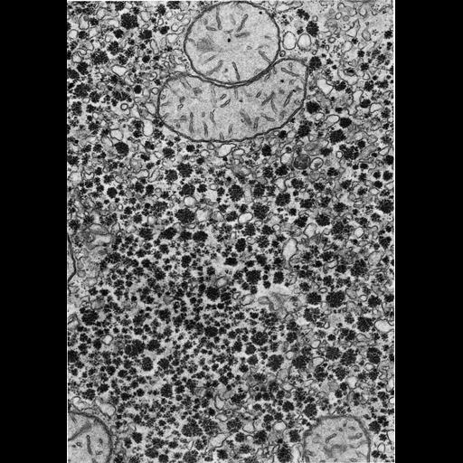
Figure 349 from Chapter 15 (Cytoplasmic Inclusions) of 'The Cell, 2nd Ed.' by Don W. Fawcett M.D. Electron micrograph of an hepatic cell from a fasted hamster after an injection of cortisone. In mammalian liver, glycogen appears as coarse rosettes and tends to be associated with the smooth endoplasmic reticulum. Image by Albert Jones. A PDF copy of the accompanying chapter is available on the ASCB’s BioEDUCATE website.

Biological Sources
NCBI Organism Classification
Phodopus
Cell Type
hepatocyte
Cellular Component
smooth endoplasmic reticulum
glycogen granule
Biological Context
Biological Process
energy reserve metabolic process
Attribution
Names
Don W. Fawcett
Albert Jones
Published
ISBN 0721635849
Link
http://www.ascb.org/bioeducate/FawcettTheCell...
Citation
Digital Object Identifier (DOI)
doi:10.7295/W9CIL35972
Archival Resource Key (ARK)
ark:/b7295/w9cil35972
Grouping This image is part of a group.
Imaging
Image Type
transmission electron microscopy (TEM)
Image Mode
illumination by electrons
detection of electrons
Parameters Imaged
electron density
Source of Contrast
stain with broad specificity
Visualization Methods
osmium tetroxide
Data Qualifiers
processed data
Dimensions
Spatial Axis Image Size Pixel Size
X 895px ——
Y 1261px ——