Licensing

Pd
Public Domain: This image is in the public domain and thus free of any copyright restrictions. However, as is the norm in scientific publishing and as a matter of courtesy, any user should credit the content provider for any public or private use of this image whenever possible. Learn more
Comments: Powered by Pnyxe

Description

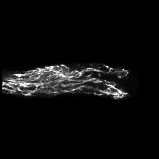
Living Lilium pollen tube labeled with dichloro-flourescein diacetate to mark the vacuole and imaged using laser scanning focal microscopy. Shown is a central 1.0 micron thick x-y slice.The growing tip is devoid of vacuole. Other images in the group reveal the distributions of additional cytoplasmic components.

Technical Details

Living lily pollen tubes were labeled and imaged with a Zeiss 510 meta confocal microscope using a 63x 1.4 NA objective lens. Image shows a central x-y plane through the tube. See Lovy-Wheeler et al. 2007 Cell Motil Cytoskel 64:217-321.

Biological Sources

NCBI Organism Classification
Lilium formosanum
(St Joseph's lily)
Cell Type
pollen tube
Cellular Component
plant-type vacuole lumen

Biological Context

Biological Process
pollen tube growth

Attribution

Names
Alenka Lovy-Wheeler
Luis Cardenas
Joseph Kunkel
Peter K Hepler

Grouping

This image is part of a group.

Imaging

Image Type
photomultiplier tube (PMT)
Imaging Mode
confocal microscopy
Parameters Imaged
fluorescence emission
Source of Contrast
compartmentalization of stain or label
Visualization Methods
carboxy-dichloroflourescein diacetate
Processing History
central x-y slice
Data Qualifiers
processed data
suitable for spatial measurements

Sample Preparation

Methods
living tissue
Relation To Intact Cell
whole mounted tissue

Dimensions

Spatial Axis Image Size Pixel Size
X 500px ——
Y 178px ——
*CIL – Cell Image Library accession number. Please use this to reference an image.