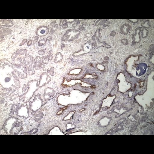
Tissue section of human prostate containing adenocarcinoma that has been immunostained for the cell-surface antigen CD133. Nuclei are stained in blue. This image is part of a large collection of images generated from numerous specimens to characterize the distribution of CD133 in human prostate tissue. A summary of the entire data set is provided below. No summary is available for CD133 immunostain of human prostate.
This image is part of a large collection of immunohistochemistry images of cell-surface antigens generated by the SCGAP Urologic Epithelial Stem Cells (UESC) Project. The overall goal of the project is to characterize and isolate epithelial stem cell populations from two urologic organs, the prostate and bladder. Links are provided below for the UESC Project database, the entire human prostate immunostain summary, the CD133 immunostain summary, and information on the specimen that this image is from. Other images of CD133 human prostate immunostains are accessible following the group link.
The "Image ID" for this image is provided in the Attribution section. Because the images are systematically named, information such as the antibody used in staining, the organism, tissue type, tissue block, and magnification are all encoded in the Image ID. As an example, the image 'CD44 98-395F HP ba 100.jpg' was stained with anti-CD44, was derived from human prostate tissue block 98-395F, is human prostate tissue (HP), and an image of microscopic field of view b within field a was captured at 100× magnification.
Complete technical details available in the Protocol Section of SCGAP UESC website (link provided). Briefly, radical prostatectomy specimens were transverse-sectioned 3mm thick and tissue blocks were embedded in precooled OCT (Tissue-tek) medium. Frozen tissue blocks were thin-sectioned (5µ) and fixed in cold acetone. A monoclonal primary antibody (PharMingen) was used followed by a biotinylated secondary antibody. Bound antibody was detected using avidin-biotin-peroxidase (Vector Labs) and diaminobenzidine (DAB). Sections were lightly counterstained with hematoxylin. Images were acquired using a digital camera and either an Olympus BX41 or Olympus BH2 microscope.
| Spatial Axis | Image Size | Pixel Size |
|---|---|---|
| X | 1600px | —— |
| Y | 1200px | —— |