Licensing
Description
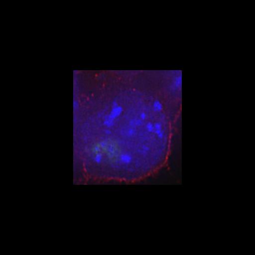
z-series image of mouse ES cells with a multi-copy alpha-globin BAC insertion. H3K9me3 (blue) marks the heterochromatic core which largely colocalizes with combined FISH signal from the lac operator repeats and BAC vector backbone (green). In contrast the BAC FISH signal (red) is more peripheral.
Technical Details
Formaldehyde fixed cells were imaged using an IMT-2 Olympus fluorescence microscope with 60x 1.4 NA objective lens. Z-series were recorded, and the slices processed by iterative constrained deconvolution. xy pixel dimensions are 0.067 microns, z spacing is 0.2 microns and number of z-slices 14. See also: P Sinclair et al. 2010 Dynamic plasticity of large scale chromatin structure revealed by self-assembly of engineered chromosome regions. J Cell Biol 109:761-776.
Biological Sources
- NCBI Organism Classification
- Mus musculus
- (house mouse)
- Cell Type
- embryonic stem cell
- Cellular Component
- heterochromatin
- nuclear chromatin
- transgene
Biological Context
- Biological Process
- nucleus organization
- chromatin organization
Attribution
- Names
- Paul Sinclair
- Qian Bian
- Matt Plutz
- Edith Heard
- Andrew Belmont
- Published
- P Sinclair et al. 2010 Dynamic plasticity of large scale chromatin structure revealed by self-assembly of engineered chromosome regions. J Cell Biol 109:761-776
- Link
- Journal of Cell Biology
- Pubmed
- 20819934
Grouping
This image is part of a group.Imaging
- Image Type
- charge coupled device (CCD)
- Imaging Mode
- fluorescence microscopy
- Parameters Imaged
- fluorescence emission
- Source of Contrast
- distribution of a specific protein
- distribution of a specific nucleic acid sequence
- Visualization Methods
- EGFP
- Processing History
- constrained iterative deconvolution
- Data Qualifiers
- processed data
- suitable for spatial measurements
Sample Preparation
- Methods
- formaldehyde fixed tissue
- Relation To Intact Cell
- whole mounted tissue
Dimensions
| Spatial Axis | Image Size | Pixel Size |
|---|---|---|
| X | 218px | 0.067μm |
| Z | 14 | 0.2μm |