Licensing
Attribution Non-Commercial Share Alike:This image is licensed under a Creative Commons Attribution, Non-Commercial Share Alike License. View License Deed | View Legal Code
Comments:
Powered by Pnyxe
Description
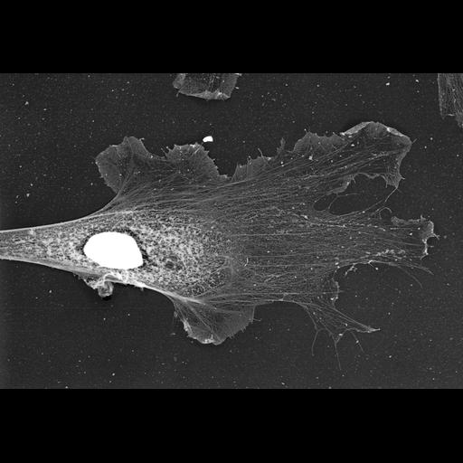
Localization of XAC (Xenopus ADF/cofilin) in Xenopus fibroblasts. Immuno-EM with XAC antibody at low magnification. High magnification view is available at CIL 24785. Nucleus and surrounding regions look bright because of high content of cellular proteins, which creates high electron density (brightness in inverted contrast). Immunogold-labeling in these central regions is very low. Image corresponds to Figure 10h from J Cell Biol. 1999 May 31;145(5):1009-26.
Technical Details
Procedures for detergent extraction, immunostaining, S1 decoration, light, and EM were described previously (Svitkina et al., 1995, 1996, 1997;Verkhovsky et al., 1995; Svitkina and Borisy, 1998).
Biological Sources
- NCBI Organism Classification
- Xenopus laevis
- (African clawed frog)
- Cell Type
- fibroblast
- Cellular Component
- lamellipodium
- Xenopus ADF/cofilin
- nucleus
Biological Context
- Biological Process
- actin filament organization
- ruffle organization
- Molecular Function
- structural constituent of cytoskeleton
Attribution
- Names
- Tatyana M. Svitkina
- Gary G. Borisy
- Published
- J Cell Biol. 1999 May 31;145(5):1009-26.
- Pubmed
- 10352018
Grouping
This image is part of a group.Imaging
- Image Type
- recorded image
- Imaging Mode
- transmission electron microscopy (TEM)
- Parameters Imaged
- optical path length gradient
- Source of Contrast
- differences in deposition of metal shadow
- Visualization Methods
- shadowing and plating
- platinum replica
- primary antibody plus labeled secondary antibody
- immuno gold
- Processing History
- unprocessed raw data
Sample Preparation
- Methods
- permeabilized tissue
- glutaraldehyde fixed tissue
- polyethylene glycol
- Relation To Intact Cell
- dispersed cell in vitro
Dimensions
| Spatial Axis | Image Size | Pixel Size |
|---|---|---|
| X | 2753px | —— |
| Y | 1878px | —— |
*CIL – Cell Image Library accession number. Please use this to reference an image.