Alternate header for print version


Licensing
Public Domain: This image is in the public domain and thus free of any copyright restrictions. However, as is the norm in scientific publishing and as a matter of courtesy, any user should credit the content provider for any public or private use of this image whenever possible. Learn more
tweet  
*CIL – Cell Image Library accession number. Please use this to reference an image.

CIL:245*  Cite 
Description

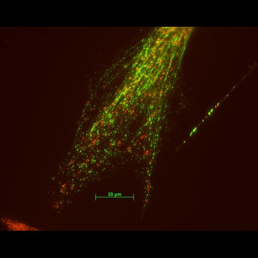
Mitochondrial distribution (visualized with Mitotracker CMXRos) was largely unaffected by partial fragmentation of the intermediate filament cytoskeleton, visualized here with vimentin antibody, after expression of a mutant form of the peripherin gene in a primary fibroblast. This primary culture derived from the mouse dorsal root ganglion was methanol-fixed at -20 degrees. Microscope - Zeiss Axioplan I Illumination - Mercury arc lamp Objective - 63X oil/1.4 NA Intermediate lens between obj and camera - 0.63X Camera - Axiocam MRM CCD Filters: 1. Ex 485/20 BP, Em 515-565 BP 2. Ex 560/40, Em 630/6

Biological Sources
NCBI Organism Classification
Mus musculus
Cell Type
fibroblast
Cellular Component
mitochondrion
type III intermediate filament
Attribution
Names
Linda Parysek
Citation
Digital Object Identifier (DOI)
doi:10.7295/W9CIL245
Archival Resource Key (ARK)
ark:/b7295/w9cil245
Imaging
Image Type
microscopy
Image Mode
fluorescence microscopy
Parameters Imaged
fluorescence emission
Source of Contrast
distribution of a specific protein
Visualization Methods
Alexa Fluor 488
CMX rosamine (Mitotracker Red)
Processing History
unprocessed raw data
Data Qualifiers
raw, unprocessed data
suitable for spatial measurements
Sample Preparation
Methods
methanol fixed tissue
Relation To Intact Cell
dispersed cells in vitro
Dimensions
Spatial Axis Image Size Pixel Size
X 1300px 104nm
Y 1030px 104nm