Licensing
Description
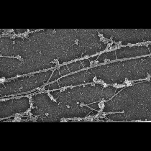
MAP4 is not required for plectin binding to microtubules (MTs). Electron microscopy of control human 356 fibroblasts (CIL:23051) and MAP4 immunodepleted (CIL:23052, this image) cells after staining with antibody to NH 2 terminus of MAP4. Cells were microinjected with an antibody to the COOH-terminal domain of MAP4 to remove MAP4 from MTs. 10-nm gold particles decorate MTs in control cells, but are absent in injected cells. Plectin forms bridges between intermediate filaments and MTs in both cells. Electron microscopy of cytoskeletons was performed as described (Svitkina et al., 1995). Briefly, cells on coverslips were lysed as for light microscopy, treated, with recombinant gelsolin NHz-terminal domain, fixed with glutaraldehyde, tannic acid and uranyl acetate, critical point dried, and coated with platinum and carbon. Image corresponds to Fig 9d in J Cell Biol. 1996 Nov;135(4):991-1007.
Biological Sources
- NCBI Organism Classification
- Homo sapiens
- (human)
- Cell Type
- fibroblast
- Cell Line
- human 356 fibroblasts
- Cellular Component
- intermediate filament
- microtubule
- plectin
Biological Context
- Biological Process
- MAP4 depletion
Attribution
- Names
- Tatyana M. Svitkina
- Alexander B. Verkhovsky,
- Gary G. Borisy
- Published
- J Cell Biol. 1996 Nov;135(4):991-1007
- Pubmed
- 8922382
Grouping
This image is part of a group.Imaging
- Image Type
- recorded image
- Imaging Mode
- transmission electron microscopy (TEM)
- Parameters Imaged
- elastic scattering of electrons
- Source of Contrast
- differences in deposition of metal shadow
- Visualization Methods
- uranyl salt
- shadowing and plating
- primary antibody plus labeled secondary antibody
- immunogold
- Processing History
- unprocessed raw data
Sample Preparation
- Methods
- glutaraldehyde fixed tissue
- tannic acid
- critical_point dried specimen
- Relation To Intact Cell
- platinum replica
Dimensions
| Spatial Axis | Image Size | Pixel Size |
|---|---|---|
| X | 4271px | —— |
| Y | 2658px | —— |