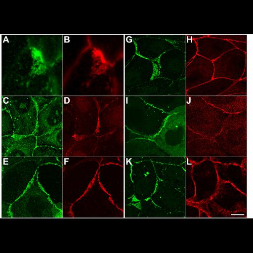
Transmembrane proteins of the tight junction (occludin, claudin-2, JAM-A), but not cytosolic proteins (cingulin, ZO-2, alpha-catenin), are incorporated into the ectopic strands formed by delta-U6 constructs. MDCK cells expressing delta-U6 (Supplementary Figures) or myc-ZNG (aa 1-806) were plated at low density in DOX-free media, fixed after 4 d and stained with antibodies against c-myc (B, D, F, H, J, and L) and antisera recognizing the transmembrane proteins occludin (A), claudin-2 (C), and JAM-A (E) or the cytosolic proteins cingulin (G), ZO-2 (I), and alpha-catenin (K). The images shown are maximum projections of 2.5–5.0-µm confocal stacks. Bar, 10 µm.
MDCK-II tet off cells were plated on glass coverslips at a low density (1.0 x 10[4] cells/ml) and incubated in the absence of doxycyclin for 4 d before processing. Cells were fixed for 30 min in ethanol on ice, permeabilized in 0.2% Triton X-100 and incubated in mouse anti-myc 9E10 or rabbit anti-myc (MBL International;1:2,500), and mouse anti-ZO-2 3E8D9 (Zymed; 1:500), rabbit anti-cingulin (Sandra Citi, Geneva; 1:500), mouse anti-occludin (Zymed; 1:300), mouse anti-claudin-2 (Zymed; 1:200), rabbit anti-canine JAM-1 (Klaus Ebnet, Munster; 1:400) and α-catenin (Sigma-Aldrich; 1:2000) followed by Cy2, Cy3 or Cy5-conjugated secondary antibodies. Cells were mounted in Mowiol with 1.0% n-propyl gallate. Confocal images were acquired on a Zeiss LSM510 Meta using a 100x Plan Apo lens (Thornwood, NY). Confocal Stacks (E,F,G,H,M,N) and image projections were generated with Zeiss LSM Image Browser version 3.2. Contrast adjustment and montages were generated using Adobe Photoshop (version 6.0; San Jose, CA). Figure 9 in Mol Biol Cell (2006). 18:721-731.