Licensing
Description
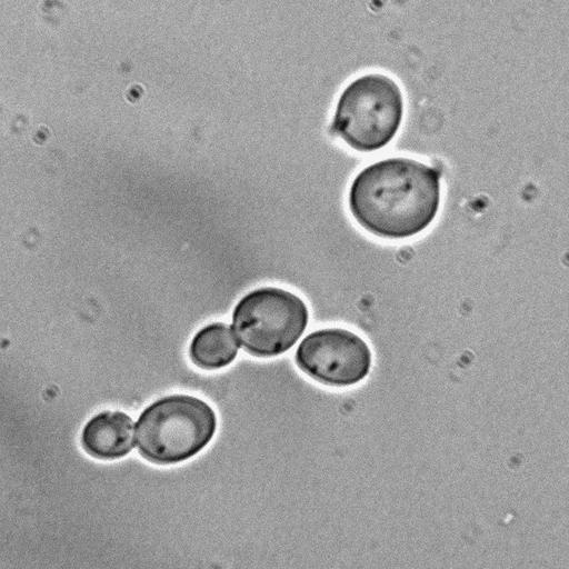
DIC image of cells constitutively expressing Tem1 localized to both spindle pole bodies (SPBs) and expressing Kin4 from the GAL1-10 promoter growing in medium with glucose. This is the control image for CIL# 13896 in which constitutive loading of Tem1 to SPBs allowed cells to progress through the cell cycle despite Kin4 overexpression. Image is Fig 5C bottom left panel in J Cell Biol. (2011) 192: 599-614. Other images in Fig 5 include CIL# 13895, 13894, 13897, 13896.
Technical Details
Cells (MATa tem1::KanMX ura3::eGFP-CNM67–TEM1::URA3 kin4::TRP1-pGAL1-10-KIN4) were grown in YPD (containing glucose). Imaging was performed at 25C using a Leica DM6000 microscope equipped with a 100x/1.40 NA oil immersion objective lens, and a digital CCD camera (DFC350, Leica). Pictures were processed with LAS AF (leica) and ImageJ software.
Biological Sources
- NCBI Organism Classification
- Saccharomyces cerevisiae
- (Baker's Yeast, Brewer's Yeast)
- Cell Line
- W303
- Cellular Component
- spindle pole body
Biological Context
- Biological Process
- mitotic cell cycle spindle orientation checkpoint
- protein phosphorylation
- regulation of exit from mitosis
- small GTPase mediated signal transduction
- mitosis
- cell division
- Molecular Function
- ATP binding
- protein serine/threonine kinase activity
- GTPase activity
- nucleotide binding
- protein binding
Attribution
- Names
- Mauricio Valerio-Santiago
- Fernando Monje-Casas
- Published
- J Cell Biol. (2011) 192: 599-614.
- Link
- http://jcb-dataviewer.rupress.org/jcb/...
- Pubmed
- 21321099
Grouping
This image is part of a group.Imaging
- Image Type
- recorded image
- Imaging Mode
- differential interference contrast microscopy
- Parameters Imaged
- optical path length gradient
- Source of Contrast
- boundaries between regions with different refractive index
- Processing History
- unprocessed raw data
- Data Qualifiers
- raw, unprocessed data
- suitable for spatial measurements
Sample Preparation
- Methods
- living tissue
- Relation To Intact Cell
- whole mounted tissue
Dimensions
| Spatial Axis | Image Size | Pixel Size |
|---|---|---|
| X | 621px | .0644μm |
| Y | 621px | .0644μm |