Licensing
Description
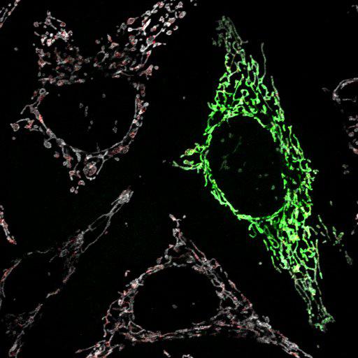
Mitochondria in HeLa cells are identified by mito-YFP (green). When fixed cells are permeabilized with 0.005% digitonin, the antibody to cyctochrome c (red), a mitochondrial matrix marker, is unable to reach and bind cytochrome c, but the antibody to Tom20 (white), a mitochondrial outer membrane marker, is now able to bind Tom20. Mito-YFP transfected HeLa cells were treated with DMSO for 3 hrs, fixed with 4% paraformaldehyde and permeabilized with 0.005% digitonin/PBS. Primary antibodies used were: anti-Cyt c mAb (BD) and anti-Tom20 mAb (BD); secondary antibodies used were: Alexa Fluor 594 and 647. Imaging was performed on an LSM510 Meta (Carl Zeiss, Inc) with a 63x 1.4 NA oil differential interference contrast Plan Apo objective. Image contrast and brightness were adjusted in the LSM image browser (Zeiss). This image corresponds to Fig 4b, 2nd row from top and is part of a differential permeabilization assay with and without the mitochondrial depolarizing agent CCCP (carbonyl cyanide m-chlorophenyl hydrazone) that is further described in Fig 4b of J Cell Biol, 191: 933-942, 2010. Images in Fig 4 include CIL#s 13733, 13734, 13729, 13730, 13731, 13732, 13717, 13718, 13719, 13720, 13721, 13722, 13723, 13724.
Biological Sources
- NCBI Organism Classification
- Homo sapiens
- (human)
- Cell Type
- endothelial cell
- Cell Line
- HeLa
- cervical carcinoma
- Cellular Component
- mitochondrion
- mitochondrial outer membrane translocase complex
- mitochondrial intermembrane space
Biological Context
- Biological Process
- respiratory electron transport chain
- protein targeting to mitochondrion
- Molecular Function
- electron transporter, transferring electrons from CoQH2-cytochrome c reductase complex and cytochrome c oxidase complex activity
- heme binding
- P-P-bond-hydrolysis-driven protein transmembrane transporter activity
Attribution
- Names
- Seok Min Jin
- Michael Lazarou
- Chunxin Wang
- Lesley A. Kane
- Derek P. Narendra
- Richard J. Youle
- Published
- J Cell Biol, 191: 933-942, 2010.
- Link
- http://jcb-dataviewer.rupress.org/jcb/...
- Pubmed
- 21115803
Grouping
This image is part of a group.Imaging
- Image Type
- recorded image
- Imaging Mode
- single-spot confocal microscopy
- Parameters Imaged
- fluorescence emission
- Source of Contrast
- distribution of a specific protein
- Visualization Methods
- EYFP
- primary antibody plus labeled secondary antibody
- Processing History
- brightness and contrast adjusted
- Data Qualifiers
- raw, unprocessed data
- suitable for spatial measurements
Sample Preparation
- Methods
- formaldehyde fixed tissue
- detergent permeabilized
- Relation To Intact Cell
- dispersed cells in vitro
Dimensions
| Spatial Axis | Image Size | Pixel Size |
|---|---|---|
| X | 512px | .1860μm |
| Y | 512px | .1860μm |