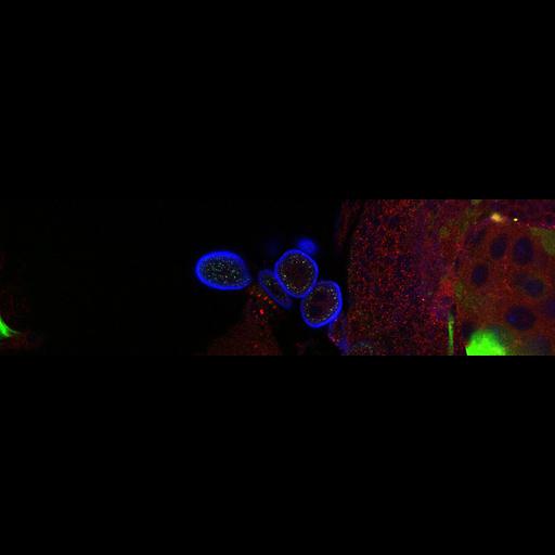
Ema localizes to endosomes. Third instar larval garland cells expressing Ema-GFP fusion protein and stained for the late endosomal and lysosomal protein Spinster (red) and plasma membrane and nuclei (blue). Ema co-localizes with some late endosomes. Wandering third instar larvae were dissected in ice-cold PBS, equilibrated in HL-3 buffer at room temperature, incubated in 0.7% HRP for 5 min at room temperature, rinsed thoroughly and fixed with Bouin’s solution for 5 min. Blocking and antibody incubation were performed in PBS containing 0.1% Triton X-100. Anti-Spinster antibody (Sweeney and Davis, 2002) used at 1:250. NeuroTrace 640/660 deep red fluorescent Nissl (Invitrogen) used at 1:500 was used to stain nuclei. Cy5-conjugated anti-HRP (Jackson Immunoresearch Laboratories) at 1:1000 was used to stain plasma membrane. Cy3-conjugated secondary antibody (Jackson Immunoresearch Laboratories, Inc.) at 1:1,000. Confocal images were acquired with a confocal microscope (model C1; Nikon) and accompanying EZ-C1 software using argon (excitation at 488 nm) and HeNe (excitation at 543 and 633 nm) lasers and a 60x Plan-Apochromat NA 1.4 objective (Nikon) at room temperature. Samples for each experiment were processed using the same confocal gain setting. Image corresponds to Fig 3B in Kim et al. J Cell Biol. 188: 717-734. 2010.
| Spatial Axis | Image Size | Pixel Size |
|---|---|---|
| X | 1024px | —— |
| Y | 288px | —— |